You are browsing environment: HUMAN GUT
CAZyme Information: MGYG000000201_02206
You are here: Home > Sequence: MGYG000000201_02206
Basic Information |
Genomic context |
Full Sequence |
Enzyme annotations |
CAZy signature domains |
CDD domains |
CAZyme hits |
PDB hits |
Swiss-Prot hits |
SignalP and Lipop annotations |
TMHMM annotations
Basic Information help
| Species | Blautia_A sp900066145 | |||||||||||
|---|---|---|---|---|---|---|---|---|---|---|---|---|
| Lineage | Bacteria; Firmicutes_A; Clostridia; Lachnospirales; Lachnospiraceae; Blautia_A; Blautia_A sp900066145 | |||||||||||
| CAZyme ID | MGYG000000201_02206 | |||||||||||
| CAZy Family | GH3 | |||||||||||
| CAZyme Description | hypothetical protein | |||||||||||
| CAZyme Property |
|
|||||||||||
| Genome Property |
|
|||||||||||
| Gene Location | Start: 30973; End: 32421 Strand: + | |||||||||||
CDD Domains download full data without filtering help
| Cdd ID | Domain | E-Value | qStart | qEnd | sStart | sEnd | Domain Description |
|---|---|---|---|---|---|---|---|
| cd19133 | AKR_AKR5F1 | 8.27e-100 | 278 | 443 | 90 | 255 | the AKR5F family of aldo-keto reductase (AKR). Klebsiella sp. 2,5-diketo-D-gluconic acid reductase (2,5-DKG reductase) is a founding member of aldo-keto reductase family 5 member F1 (AKR5F1). It catalyzes the reduction of 2,5-diketo-D-gluconic acid (25DKG) to 2-keto-L-gulonic acid (2KLG). |
| cd19071 | AKR_AKR1-5-like | 1.29e-79 | 276 | 439 | 79 | 250 | AKR1/2/3/4/5 family of aldo-keto reductase (AKR) and similar proteins. Aldo-keto reductases (AKRs) are a superfamily of soluble NAD(P)(H) oxidoreductases whose chief purpose is to reduce aldehydes and ketones to primary and secondary alcohols. The family includes AKR1A/B/C/D/E/G/I, AKR2A/B/C/D/E, AKR3A/B/C/D/E/G, AKR4A/B/C, AKR5A/B/C/D/E/F/G/H, and similar proteins. |
| COG0656 | ARA1 | 1.39e-78 | 279 | 456 | 96 | 276 | Aldo/keto reductase, related to diketogulonate reductase [Secondary metabolites biosynthesis, transport and catabolism]. |
| cd19157 | AKR_AKR5G1-3 | 5.49e-69 | 278 | 444 | 91 | 256 | AKR5G family of aldo-keto reductase (AKR). Bacillus subtilis glyoxal reductase (GR), uncharacterized oxidoreductase YtbE, and Bacillus aryabhattai aldo-keto reductase are founding members of aldo-keto reductase family 5 member G1-3 (AKR5G1-3), respectively. GR (YvgN, EC 1.1.1.283), also called methylglyoxal reductase, reduces glyoxal and methylglyoxal (2-oxopropanal). It is not involved in vitamin B6 biosynthesis. |
| cd19131 | AKR_AKR5C2 | 5.81e-67 | 273 | 443 | 86 | 256 | Escherichia coli 2,5-diketo-D-gluconic acid reductase A (DkgA/YqhE) and similar proteins. Escherichia coli DkgA/YqhE is a founding member of aldo-keto reductase family 5 member C2 (AKR5C2). DkgA/YqhE (EC 1.1.1.274), also called 2,5-DKG reductase A, or 2,5-DKGR A, or 25DKGR-A, or AKR5C, catalyzes the reduction of 2,5-diketo-D-gluconic acid (25DKG) to 2-keto-L-gulonic acid (2KLG). It is also capable of stereoselective -keto ester reductions on ethyl acetoacetate and other 2-substituted derivatives. |
CAZyme Hits help
| Hit ID | E-Value | Query Start | Query End | Hit Start | Hit End |
|---|---|---|---|---|---|
| AFN84568.1 | 6.61e-137 | 1 | 275 | 267 | 543 |
| ANY69911.1 | 5.53e-116 | 1 | 277 | 170 | 450 |
| QAY32068.1 | 8.93e-93 | 1 | 275 | 173 | 432 |
| QOL32359.1 | 2.83e-89 | 1 | 275 | 173 | 435 |
| QOL35032.1 | 2.83e-89 | 1 | 275 | 173 | 435 |
PDB Hits download full data without filtering help
| Hit ID | E-Value | Query Start | Query End | Hit Start | Hit End | Description |
|---|---|---|---|---|---|---|
| 4MHB_A | 1.20e-68 | 278 | 463 | 107 | 291 | Structureof a putative reductase from Yersinia pestis [Yersinia pestis],4MHB_B Structure of a putative reductase from Yersinia pestis [Yersinia pestis],4MHB_C Structure of a putative reductase from Yersinia pestis [Yersinia pestis],4MHB_D Structure of a putative reductase from Yersinia pestis [Yersinia pestis],4MHB_E Structure of a putative reductase from Yersinia pestis [Yersinia pestis],4MHB_F Structure of a putative reductase from Yersinia pestis [Yersinia pestis] |
| 1VP5_A | 9.58e-64 | 276 | 465 | 108 | 296 | Crystalstructure of 2,5-diketo-D-gluconic acid reductase (TM1009) from Thermotoga maritima at 2.40 A resolution [Thermotoga maritima MSB8],1VP5_B Crystal structure of 2,5-diketo-D-gluconic acid reductase (TM1009) from Thermotoga maritima at 2.40 A resolution [Thermotoga maritima MSB8] |
| 3D3F_A | 4.82e-49 | 182 | 443 | 15 | 261 | CrystalStructure of Yvgn and cofactor NADPH from Bacillus subtilis [Bacillus subtilis],3D3F_B Crystal Structure of Yvgn and cofactor NADPH from Bacillus subtilis [Bacillus subtilis] |
| 3F7J_A | 4.95e-49 | 182 | 443 | 16 | 262 | B.subtilisYvgN [Bacillus subtilis],3F7J_B B.subtilis YvgN [Bacillus subtilis] |
| 4F40_A | 1.96e-47 | 270 | 444 | 94 | 275 | X-raycrystal structure of Apo prostaglandin f synthase from Leishmania major Friedlin [Leishmania major],4F40_B X-ray crystal structure of Apo prostaglandin f synthase from Leishmania major Friedlin [Leishmania major],4G5D_A X-ray crystal structure of Prostaglandin f synthase from Leishmania major Friedlin bound to NADPH [Leishmania major strain Friedlin],4G5D_B X-ray crystal structure of Prostaglandin f synthase from Leishmania major Friedlin bound to NADPH [Leishmania major strain Friedlin] |
Swiss-Prot Hits download full data without filtering help
| Hit ID | E-Value | Query Start | Query End | Hit Start | Hit End | Description |
|---|---|---|---|---|---|---|
| O32210 | 2.71e-48 | 182 | 443 | 16 | 262 | Glyoxal reductase OS=Bacillus subtilis (strain 168) OX=224308 GN=yvgN PE=1 SV=1 |
| P22045 | 9.64e-47 | 270 | 444 | 90 | 271 | 9,11-endoperoxide prostaglandin H2 reductase OS=Leishmania major OX=5664 GN=P100/11E PE=1 SV=3 |
| Q4DJ07 | 1.41e-44 | 279 | 443 | 98 | 264 | 9,11-endoperoxide prostaglandin H2 reductase OS=Trypanosoma cruzi (strain CL Brener) OX=353153 GN=Tc00.1047053511287.49 PE=1 SV=2 |
| Q8XBT6 | 4.44e-43 | 275 | 443 | 90 | 261 | 2,5-diketo-D-gluconic acid reductase A OS=Escherichia coli O157:H7 OX=83334 GN=dkgA PE=3 SV=1 |
| Q46857 | 4.44e-43 | 275 | 443 | 90 | 261 | 2,5-diketo-D-gluconic acid reductase A OS=Escherichia coli (strain K12) OX=83333 GN=dkgA PE=1 SV=3 |
SignalP and Lipop Annotations help
This protein is predicted as OTHER
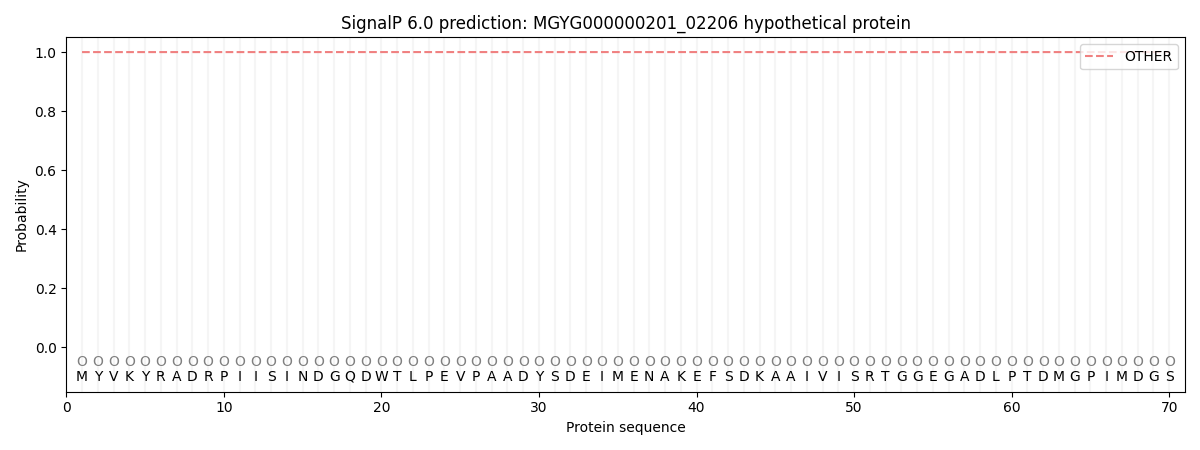
| Other | SP_Sec_SPI | LIPO_Sec_SPII | TAT_Tat_SPI | TATLIP_Sec_SPII | PILIN_Sec_SPIII |
|---|---|---|---|---|---|
| 1.000060 | 0.000001 | 0.000000 | 0.000000 | 0.000000 | 0.000000 |