You are browsing environment: HUMAN GUT
CAZyme Information: MGYG000003965_00831
You are here: Home > Sequence: MGYG000003965_00831
Basic Information |
Genomic context |
Full Sequence |
Enzyme annotations |
CAZy signature domains |
CDD domains |
CAZyme hits |
PDB hits |
Swiss-Prot hits |
SignalP and Lipop annotations |
TMHMM annotations
Basic Information help
| Species | ||||||||||||
|---|---|---|---|---|---|---|---|---|---|---|---|---|
| Lineage | Bacteria; Verrucomicrobiota; Lentisphaeria; Victivallales; Victivallaceae; UMGS1518; | |||||||||||
| CAZyme ID | MGYG000003965_00831 | |||||||||||
| CAZy Family | GT112 | |||||||||||
| CAZyme Description | hypothetical protein | |||||||||||
| CAZyme Property |
|
|||||||||||
| Genome Property |
|
|||||||||||
| Gene Location | Start: 1295; End: 2731 Strand: - | |||||||||||
CAZyme Signature Domains help
| Family | Start | End | Evalue | family coverage |
|---|---|---|---|---|
| GT112 | 66 | 438 | 2.8e-165 | 0.9946524064171123 |
CDD Domains download full data without filtering help
| Cdd ID | Domain | E-Value | qStart | qEnd | sStart | sEnd | Domain Description |
|---|---|---|---|---|---|---|---|
| TIGR04414 | hepto_Aah_TibC | 0.0 | 63 | 439 | 1 | 374 | autotransporter strand-loop-strand O-heptosyltransferase. Both Aah (autotransporter adhesin heptosyltransferase) and TibC (tib is enterotoxigenic invasion locus B) are protein O-heptosyltransferases that act on multiple sites from repeat regions of proteins exported by autotransporters. Aah glycosylates AIDA, or autotransporter adhesin involved in diffuse adherence, TibC acts on TibA, but TibC can replace Aah. [Protein fate, Protein modification and repair] |
| cd03789 | GT9_LPS_heptosyltransferase | 1.81e-19 | 169 | 438 | 18 | 277 | lipopolysaccharide heptosyltransferase and similar proteins. Lipopolysaccharide heptosyltransferase (2.4.99.B6) is involved in the biosynthesis of lipooligosaccharide (LOS). Lipopolysaccharide (LPS) is a major component of the outer membrane of gram-negative bacteria. LPS heptosyltransferase transfers heptose molecules from ADP-heptose to 3-deoxy-D-manno-octulosonic acid (KDO), a part of the inner core component of LPS. This family also contains lipopolysaccharide 1,2-N-acetylglucosaminetransferase EC 2.4.1.56 and belongs to the GT-B structural superfamily of glycoslytransferases, which have characteristic N- and C-terminal domains each containing a typical Rossmann fold. The two domains have high structural homology despite minimal sequence homology. The large cleft that separates the two domains includes the catalytic center and permits a high degree of flexibility. |
| pfam01075 | Glyco_transf_9 | 5.88e-07 | 248 | 384 | 76 | 220 | Glycosyltransferase family 9 (heptosyltransferase). Members of this family belong to glycosyltransferase family 9. Lipopolysaccharide is a major component of the outer leaflet of the outer membrane in Gram-negative bacteria. It is composed of three domains; lipid A, Core oligosaccharide and the O-antigen. All of these enzymes transfer heptose to the lipopolysaccharide core. |
CAZyme Hits help
| Hit ID | E-Value | Query Start | Query End | Hit Start | Hit End |
|---|---|---|---|---|---|
| AVM45539.1 | 8.80e-214 | 36 | 443 | 26 | 425 |
| AVM43209.1 | 1.26e-202 | 51 | 442 | 36 | 427 |
| CAL79844.1 | 2.90e-160 | 64 | 442 | 54 | 431 |
| AVM43989.1 | 5.86e-160 | 51 | 439 | 21 | 410 |
| QDQ85661.1 | 1.06e-154 | 55 | 440 | 28 | 418 |
PDB Hits download full data without filtering help
| Hit ID | E-Value | Query Start | Query End | Hit Start | Hit End | Description |
|---|---|---|---|---|---|---|
| 4RB4_A | 7.56e-142 | 63 | 439 | 13 | 386 | Crystalstructure of dodecameric iron-containing heptosyltransferase TibC in complex with ADP-D-beta-D-heptose at 3.9 angstrom resolution [Escherichia coli DEC13E],4RB4_B Crystal structure of dodecameric iron-containing heptosyltransferase TibC in complex with ADP-D-beta-D-heptose at 3.9 angstrom resolution [Escherichia coli DEC13E],4RB4_C Crystal structure of dodecameric iron-containing heptosyltransferase TibC in complex with ADP-D-beta-D-heptose at 3.9 angstrom resolution [Escherichia coli DEC13E],4RB4_D Crystal structure of dodecameric iron-containing heptosyltransferase TibC in complex with ADP-D-beta-D-heptose at 3.9 angstrom resolution [Escherichia coli DEC13E],4RB4_E Crystal structure of dodecameric iron-containing heptosyltransferase TibC in complex with ADP-D-beta-D-heptose at 3.9 angstrom resolution [Escherichia coli DEC13E],4RB4_F Crystal structure of dodecameric iron-containing heptosyltransferase TibC in complex with ADP-D-beta-D-heptose at 3.9 angstrom resolution [Escherichia coli DEC13E],4RB4_G Crystal structure of dodecameric iron-containing heptosyltransferase TibC in complex with ADP-D-beta-D-heptose at 3.9 angstrom resolution [Escherichia coli DEC13E],4RB4_H Crystal structure of dodecameric iron-containing heptosyltransferase TibC in complex with ADP-D-beta-D-heptose at 3.9 angstrom resolution [Escherichia coli DEC13E],4RB4_I Crystal structure of dodecameric iron-containing heptosyltransferase TibC in complex with ADP-D-beta-D-heptose at 3.9 angstrom resolution [Escherichia coli DEC13E],4RB4_J Crystal structure of dodecameric iron-containing heptosyltransferase TibC in complex with ADP-D-beta-D-heptose at 3.9 angstrom resolution [Escherichia coli DEC13E],4RB4_K Crystal structure of dodecameric iron-containing heptosyltransferase TibC in complex with ADP-D-beta-D-heptose at 3.9 angstrom resolution [Escherichia coli DEC13E],4RB4_L Crystal structure of dodecameric iron-containing heptosyltransferase TibC in complex with ADP-D-beta-D-heptose at 3.9 angstrom resolution [Escherichia coli DEC13E] |
| 4RAP_A | 3.48e-140 | 63 | 439 | 13 | 386 | Crystalstructure of bacterial iron-containing dodecameric glycosyltransferase TibC from enterotoxigenic E.coli H10407 [Escherichia coli ETEC H10407],4RAP_B Crystal structure of bacterial iron-containing dodecameric glycosyltransferase TibC from enterotoxigenic E.coli H10407 [Escherichia coli ETEC H10407],4RAP_C Crystal structure of bacterial iron-containing dodecameric glycosyltransferase TibC from enterotoxigenic E.coli H10407 [Escherichia coli ETEC H10407],4RAP_D Crystal structure of bacterial iron-containing dodecameric glycosyltransferase TibC from enterotoxigenic E.coli H10407 [Escherichia coli ETEC H10407],4RAP_E Crystal structure of bacterial iron-containing dodecameric glycosyltransferase TibC from enterotoxigenic E.coli H10407 [Escherichia coli ETEC H10407],4RAP_F Crystal structure of bacterial iron-containing dodecameric glycosyltransferase TibC from enterotoxigenic E.coli H10407 [Escherichia coli ETEC H10407],4RAP_G Crystal structure of bacterial iron-containing dodecameric glycosyltransferase TibC from enterotoxigenic E.coli H10407 [Escherichia coli ETEC H10407],4RAP_H Crystal structure of bacterial iron-containing dodecameric glycosyltransferase TibC from enterotoxigenic E.coli H10407 [Escherichia coli ETEC H10407],4RAP_I Crystal structure of bacterial iron-containing dodecameric glycosyltransferase TibC from enterotoxigenic E.coli H10407 [Escherichia coli ETEC H10407],4RAP_J Crystal structure of bacterial iron-containing dodecameric glycosyltransferase TibC from enterotoxigenic E.coli H10407 [Escherichia coli ETEC H10407],4RAP_K Crystal structure of bacterial iron-containing dodecameric glycosyltransferase TibC from enterotoxigenic E.coli H10407 [Escherichia coli ETEC H10407],4RAP_L Crystal structure of bacterial iron-containing dodecameric glycosyltransferase TibC from enterotoxigenic E.coli H10407 [Escherichia coli ETEC H10407] |
Swiss-Prot Hits download full data without filtering help
| Hit ID | E-Value | Query Start | Query End | Hit Start | Hit End | Description |
|---|---|---|---|---|---|---|
| Q9S4K6 | 2.55e-142 | 63 | 439 | 13 | 386 | Glycosyltransferase TibC OS=Escherichia coli O78:H11 (strain H10407 / ETEC) OX=316401 GN=tibC PE=1 SV=1 |
SignalP and Lipop Annotations help
This protein is predicted as OTHER
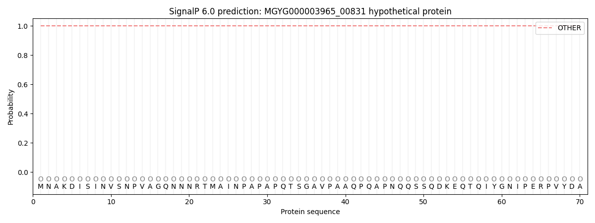
| Other | SP_Sec_SPI | LIPO_Sec_SPII | TAT_Tat_SPI | TATLIP_Sec_SPII | PILIN_Sec_SPIII |
|---|---|---|---|---|---|
| 1.000063 | 0.000000 | 0.000000 | 0.000000 | 0.000000 | 0.000000 |