You are browsing environment: HUMAN GUT
CAZyme Information: MGYG000004341_00773
You are here: Home > Sequence: MGYG000004341_00773
Basic Information |
Genomic context |
Full Sequence |
Enzyme annotations |
CAZy signature domains |
CDD domains |
CAZyme hits |
PDB hits |
Swiss-Prot hits |
SignalP and Lipop annotations |
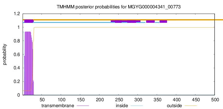
TMHMM annotations
Basic Information help
| Species | CAG-110 sp000435995 | |||||||||||
|---|---|---|---|---|---|---|---|---|---|---|---|---|
| Lineage | Bacteria; Firmicutes_A; Clostridia; Oscillospirales; Oscillospiraceae; CAG-110; CAG-110 sp000435995 | |||||||||||
| CAZyme ID | MGYG000004341_00773 | |||||||||||
| CAZy Family | GH24 | |||||||||||
| CAZyme Description | hypothetical protein | |||||||||||
| CAZyme Property |
|
|||||||||||
| Genome Property |
|
|||||||||||
| Gene Location | Start: 37206; End: 38708 Strand: - | |||||||||||
CAZyme Signature Domains help
| Family | Start | End | Evalue | family coverage |
|---|---|---|---|---|
| GH24 | 271 | 413 | 5.3e-23 | 0.9854014598540146 |
CDD Domains download full data without filtering help
| Cdd ID | Domain | E-Value | qStart | qEnd | sStart | sEnd | Domain Description |
|---|---|---|---|---|---|---|---|
| cd00737 | lyz_endolysin_autolysin | 2.24e-27 | 275 | 418 | 2 | 136 | endolysin and autolysin. The dsDNA phages of eubacteria use endolysins or muralytic enzymes in conjunction with hollin, a small membrane protein, to degrade the peptidoglycan found in bacterial cell walls. Similarly, bacteria produce autolysins to facilitate the biosynthesis of its cell wall heteropolymer peptidoglycan and cell division. Endolysins and autolysins are found in viruses and bacteria, respectively. Both endolysin and autolysin enzymes cleave the glycosidic beta 1,4-bonds between the N-acetylmuramic acid and the N-acetylglucosamine of the peptidoglycan. |
| COG3772 | RrrD | 2.95e-14 | 271 | 422 | 8 | 152 | Phage-related lysozyme (muramidase), GH24 family [Cell wall/membrane/envelope biogenesis]. |
| cd16901 | lyz_P1 | 1.16e-09 | 269 | 413 | 1 | 135 | P1 lysozyme Lyz-like proteins. Enterobacteria phage P1 lysozyme Lyz is secreted to the Escherichia coli periplasm where it is membrane bound and inactive. Activation involves the release from the membrane, an intramolecular thiol-disulfide isomerization and extensive structural rearrangement of the N-terminal region. The dsDNA phages of eubacteria use endolysins or muralytic enzymes in conjunction with hollin, a small membrane protein, to degrade the peptidoglycan found in bacterial cell walls. Similarly, bacteria produce autolysins to facilitate the biosynthesis of its cell wall heteropolymer peptidoglycan and cell division. Endolysins and autolysins are found in viruses and bacteria, respectively. Both endolysin and autolysin enzymes cleave the glycosidic beta 1,4-bonds between the N-acetylmuramic acid and the N-acetylglucosamine of the peptidoglycan. |
| pfam00395 | SLH | 2.52e-09 | 80 | 121 | 1 | 42 | S-layer homology domain. |
| NF033190 | inl_like_NEAT_1 | 2.04e-08 | 89 | 186 | 651 | 749 | NEAT domain-containing leucine-rich repeat protein. Members of this family have an N-terminal NEAT (near transporter) domain often associated with iron transport, followed by a leucine-rich repeat region with significant sequence similarity to the internalins of Listeria monocytogenes. However, since Bacillus cereus (from which this protein was described, in PMID:16978259) is not considered an intracellular pathogen, and the function may be iron transport rather than internalization, applying the name "internalin" to this family probably would be misleading. |
CAZyme Hits help
| Hit ID | E-Value | Query Start | Query End | Hit Start | Hit End |
|---|---|---|---|---|---|
| BCK83697.1 | 4.13e-36 | 79 | 258 | 352 | 526 |
| QCI60723.1 | 2.24e-35 | 79 | 263 | 392 | 571 |
| ARE59829.1 | 5.80e-34 | 78 | 255 | 783 | 957 |
| QQR06511.1 | 5.80e-34 | 78 | 255 | 783 | 957 |
| QNL43287.1 | 1.35e-30 | 54 | 257 | 361 | 553 |
PDB Hits download full data without filtering help
| Hit ID | E-Value | Query Start | Query End | Hit Start | Hit End | Description |
|---|---|---|---|---|---|---|
| 6BT4_A | 5.56e-09 | 80 | 259 | 26 | 196 | Crystalstructure of the SLH domain of Sap from Bacillus anthracis in complex with a pyruvylated SCWP unit [Bacillus anthracis] |
| 3PYW_A | 5.65e-09 | 80 | 259 | 5 | 175 | Thestructure of the SLH domain from B. anthracis surface array protein at 1.8A [Bacillus anthracis] |
| 6ET6_A | 8.54e-07 | 236 | 418 | 20 | 194 | ChainA, Lysozyme [Acinetobacter baumannii] |
| 7M5I_A | 2.25e-06 | 271 | 418 | 10 | 154 | ChainA, Endolysin [Escherichia coli O157 typing phage 15],7M5I_B Chain B, Endolysin [Escherichia coli O157 typing phage 15] |
Swiss-Prot Hits download full data without filtering help
| Hit ID | E-Value | Query Start | Query End | Hit Start | Hit End | Description |
|---|---|---|---|---|---|---|
| C6CRV0 | 5.61e-18 | 70 | 258 | 1273 | 1458 | Endo-1,4-beta-xylanase A OS=Paenibacillus sp. (strain JDR-2) OX=324057 GN=xynA1 PE=1 SV=1 |
| P19424 | 8.29e-18 | 68 | 258 | 29 | 213 | Endoglucanase OS=Bacillus sp. (strain KSM-635) OX=1415 PE=1 SV=1 |
| P38535 | 4.11e-14 | 72 | 258 | 900 | 1080 | Exoglucanase XynX OS=Acetivibrio thermocellus OX=1515 GN=xynX PE=3 SV=1 |
| P38536 | 4.75e-14 | 61 | 258 | 1663 | 1854 | Amylopullulanase OS=Thermoanaerobacterium thermosulfurigenes OX=33950 GN=amyB PE=3 SV=2 |
| P38537 | 1.03e-10 | 75 | 257 | 28 | 204 | Surface-layer 125 kDa protein OS=Lysinibacillus sphaericus OX=1421 PE=3 SV=1 |
SignalP and Lipop Annotations help
This protein is predicted as SP
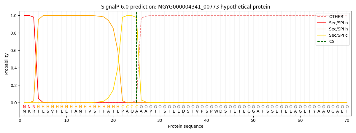
| Other | SP_Sec_SPI | LIPO_Sec_SPII | TAT_Tat_SPI | TATLIP_Sec_SPII | PILIN_Sec_SPIII |
|---|---|---|---|---|---|
| 0.000214 | 0.999147 | 0.000168 | 0.000171 | 0.000155 | 0.000137 |