You are browsing environment: HUMAN GUT
CAZyme Information: MGYG000000944_00272
You are here: Home > Sequence: MGYG000000944_00272
Basic Information |
Genomic context |
Full Sequence |
Enzyme annotations |
CAZy signature domains |
CDD domains |
CAZyme hits |
PDB hits |
Swiss-Prot hits |
SignalP and Lipop annotations |
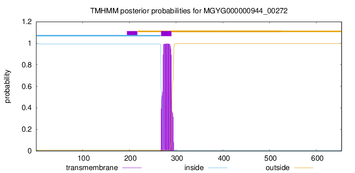
TMHMM annotations
Basic Information help
| Species | CAG-1427 sp900538545 | |||||||||||
|---|---|---|---|---|---|---|---|---|---|---|---|---|
| Lineage | Bacteria; Actinobacteriota; Coriobacteriia; Coriobacteriales; Eggerthellaceae; CAG-1427; CAG-1427 sp900538545 | |||||||||||
| CAZyme ID | MGYG000000944_00272 | |||||||||||
| CAZy Family | CE4 | |||||||||||
| CAZyme Description | hypothetical protein | |||||||||||
| CAZyme Property |
|
|||||||||||
| Genome Property |
|
|||||||||||
| Gene Location | Start: 63086; End: 65050 Strand: - | |||||||||||
Full Sequence Download help
| MPHDYRPAHH QHDHARPHRR KPQRRTSNRE TVQSDELSST NGIRMVKDAQ SFNHDRNPHL | 60 |
| SQTSLPTIGA VGTPHPKSPA ARAMQQQARP GQEAPGFGVE EAPFESTGVR THGAGNPLYT | 120 |
| GETYPVRGTH TDGSLPPVHA HDQQTNASFT GSHRATQAFS SEYPATHASQ SSRFRTVPPT | 180 |
| SDQVDPQAEQ AARSRAADTA NPRQRSYAGH SASFAAVEHA QASPQPYDYQ DYTAASGRYK | 240 |
| ASNAQAASPA NFKPLTSTSK HARKRTKAWP RVIAAVAIVV ALGIGGFFGY QQIAFAGPIT | 300 |
| ATVNGEKMTL EGSERSIEGL LDNNIVQVTP GNFVAVDESV LREGDGTRCT AIINEQPVSD | 360 |
| LNAHINEGDQ VVVSNGTDVM EDYTDSAPTI TPAGYSKAGQ YGALHVFLAG QDGETVTRTG | 420 |
| SESGKTIEQV TKEKIDNRLV YYNANSNGEK VIALTFDDGP WDGSTEQILD ILKENGAKAT | 480 |
| FYTIGEQISS HSDQIARMAN EGHEIATHTW DHAAGSGKGV SLNLMSTTER KEEVQKGMDA | 540 |
| IKDAANKDAS VYFRAPGGNF NDSVANDLRD MVQGEIGWNI DTEDWRRPGA DTIASRIKSA | 600 |
| GPGEVILMHD GGGDRSQTVA ALREALPYLK SQGYKFVTIS ELIEAYPYQE GQHS | 654 |
CAZyme Signature Domains help
| Family | Start | End | Evalue | family coverage |
|---|---|---|---|---|
| CE4 | 444 | 570 | 2.4e-28 | 0.8923076923076924 |
CDD Domains download full data without filtering help
| Cdd ID | Domain | E-Value | qStart | qEnd | sStart | sEnd | Domain Description |
|---|---|---|---|---|---|---|---|
| cd10917 | CE4_NodB_like_6s_7s | 1.43e-62 | 450 | 630 | 1 | 171 | Catalytic NodB homology domain of rhizobial NodB-like proteins. This family belongs to the large and functionally diverse carbohydrate esterase 4 (CE4) superfamily, whose members show strong sequence similarity with some variability due to their distinct carbohydrate substrates. It includes many rhizobial NodB chitooligosaccharide N-deacetylase (EC 3.5.1.-)-like proteins, mainly from bacteria and eukaryotes, such as chitin deacetylases (EC 3.5.1.41), bacterial peptidoglycan N-acetylglucosamine deacetylases (EC 3.5.1.-), and acetylxylan esterases (EC 3.1.1.72), which catalyze the N- or O-deacetylation of substrates such as acetylated chitin, peptidoglycan, and acetylated xylan. All members of this family contain a catalytic NodB homology domain with the same overall topology and a deformed (beta/alpha)8 barrel fold with 6- or 7 strands. Their catalytic activity is dependent on the presence of a divalent cation, preferably cobalt or zinc, and they employ a conserved His-His-Asp zinc-binding triad closely associated with the conserved catalytic base (aspartic acid) and acid (histidine) to carry out acid/base catalysis. Several family members show diversity both in metal ion specificities and in the residues that coordinate the metal. |
| cd10962 | CE4_GT2-like | 1.90e-59 | 450 | 643 | 1 | 195 | Catalytic NodB homology domain of uncharacterized bacterial glycosyl transferase, group 2-like family proteins. This family includes many uncharacterized bacterial proteins containing an N-terminal GH18 (glycosyl hydrolase, family 18) domain, a middle NodB-like homology domain, and a C-terminal GT2-like (glycosyl transferase group 2) domain. Although their biological function is unknown, members in this family contain a middle NodB homology domain that is similar to the catalytic domain of Streptococcus pneumoniae polysaccharide deacetylase PgdA (SpPgdA), an extracellular metal-dependent polysaccharide deacetylase with de-N-acetylase activity toward a hexamer of chitooligosaccharide N-acetylglucosamine, but not shorter chitooligosaccharides or a synthetic peptidoglycan tetrasaccharide. Like SpPgdA, this family is a member of the carbohydrate esterase 4 (CE4) superfamily. The presence of three domains suggests that members of this family may be multifunctional. |
| cd10954 | CE4_CtAXE_like | 5.72e-59 | 450 | 642 | 1 | 180 | Catalytic NodB homology domain of Clostridium thermocellum acetylxylan esterase and its bacterial homologs. This family is represented by Clostridium thermocellum acetylxylan esterase (CtAXE, EC 3.1.1.72), a member of the carbohydrate esterase 4 (CE4) superfamily. CtAXE deacetylates O-acetylated xylan, a key component of plant cell walls. It shows no detectable activity on generic esterase substrates including para-nitrophenyl acetate. It is specific for sugar-based substrates and will precipitate acetylxylan, as a consequence of deacetylation. CtAXE is a monomeric protein containing a catalytic NodB homology domain with the same overall topology and a deformed (beta/alpha)8 barrel fold as other CE4 esterases. However, due to differences in the topography of the substrate-binding groove, the chemistry of the active center, and metal ion coordination, CtAXE has different metal ion preference and lacks activity on N-acetyl substrates. It is significantly activated by Co2+. Moreover, CtAXE displays distinctly different ligand coordination to the metal ion, utilizing an aspartate, a histidine, and four water molecules, as opposed to the conserved His-His-Asp zinc-binding triad of other CE4 esterases. |
| TIGR02764 | spore_ybaN_pdaB | 1.16e-52 | 447 | 643 | 3 | 191 | polysaccharide deacetylase family sporulation protein PdaB. This model describes the YbaN protein family, also called PdaB and SpoVIE, of Gram-positive bacteria. Although ybaN null mutants have only a mild sporulation defect, ybaN/ytrI double mutants show drastically reducted sporulation efficiencies. This synthetic defect suggests the role of this sigmaE-controlled gene in sporulation had been masked by functional redundancy. Members of this family are homologous to a characterized polysaccharide deacetylase; the exact function this protein family is unknown. [Cellular processes, Sporulation and germination] |
| COG0726 | CDA1 | 2.27e-50 | 441 | 644 | 56 | 258 | Peptidoglycan/xylan/chitin deacetylase, PgdA/CDA1 family [Carbohydrate transport and metabolism, Cell wall/membrane/envelope biogenesis]. |
CAZyme Hits help
| Hit ID | E-Value | Query Start | Query End | Hit Start | Hit End |
|---|---|---|---|---|---|
| BCA89127.1 | 2.48e-109 | 272 | 643 | 161 | 528 |
| BCS56772.1 | 1.93e-108 | 272 | 643 | 161 | 528 |
| BAN76597.1 | 2.04e-108 | 272 | 643 | 163 | 530 |
| BBH49987.1 | 3.35e-106 | 262 | 646 | 96 | 482 |
| QOS70093.1 | 2.91e-99 | 279 | 647 | 1 | 363 |
PDB Hits download full data without filtering help
| Hit ID | E-Value | Query Start | Query End | Hit Start | Hit End | Description |
|---|---|---|---|---|---|---|
| 7FBW_A | 2.29e-31 | 444 | 644 | 111 | 301 | ChainA, Predicted xylanase/chitin deacetylase [Caldanaerobacter subterraneus subsp. tengcongensis MB4] |
| 2C1G_A | 2.72e-28 | 449 | 643 | 235 | 416 | Structureof Streptococcus pneumoniae peptidoglycan deacetylase (SpPgdA) [Streptococcus pneumoniae R6] |
| 6H8L_A | 9.73e-28 | 449 | 652 | 9 | 199 | Structureof peptidoglycan deacetylase PdaC from Bacillus subtilis [Bacillus subtilis subsp. subtilis str. 168],6H8L_B Structure of peptidoglycan deacetylase PdaC from Bacillus subtilis [Bacillus subtilis subsp. subtilis str. 168] |
| 2C1I_A | 1.21e-27 | 449 | 643 | 235 | 416 | Structureof Streptococcus pneumoniae peptidoglycan deacetylase (SpPgdA) D 275 N Mutant. [Streptococcus pneumoniae R6] |
| 6H8N_A | 6.24e-27 | 449 | 652 | 9 | 199 | Structureof peptidoglycan deacetylase PdaC from Bacillus subtilis - mutant D285S [Bacillus subtilis subsp. subtilis str. 168],6H8N_B Structure of peptidoglycan deacetylase PdaC from Bacillus subtilis - mutant D285S [Bacillus subtilis subsp. subtilis str. 168] |
Swiss-Prot Hits download full data without filtering help
| Hit ID | E-Value | Query Start | Query End | Hit Start | Hit End | Description |
|---|---|---|---|---|---|---|
| Q81AF4 | 9.68e-39 | 447 | 643 | 19 | 209 | Peptidoglycan-N-acetylglucosamine deacetylase BC_3618 OS=Bacillus cereus (strain ATCC 14579 / DSM 31 / CCUG 7414 / JCM 2152 / NBRC 15305 / NCIMB 9373 / NCTC 2599 / NRRL B-3711) OX=226900 GN=BC_3618 PE=1 SV=1 |
| Q52845 | 8.27e-29 | 444 | 641 | 15 | 217 | Chitooligosaccharide deacetylase OS=Mesorhizobium japonicum (strain LMG 29417 / CECT 9101 / MAFF 303099) OX=266835 GN=nodB PE=3 SV=2 |
| Q8DP63 | 2.21e-27 | 449 | 643 | 267 | 448 | Peptidoglycan-N-acetylglucosamine deacetylase OS=Streptococcus pneumoniae (strain ATCC BAA-255 / R6) OX=171101 GN=pgdA PE=1 SV=1 |
| P04675 | 6.36e-27 | 447 | 642 | 18 | 218 | Chitooligosaccharide deacetylase OS=Bradyrhizobium sp. (strain ANU 289) OX=186901 GN=nodB PE=3 SV=2 |
| P04339 | 8.04e-27 | 450 | 635 | 21 | 211 | Chitooligosaccharide deacetylase OS=Rhizobium leguminosarum bv. viciae OX=387 GN=nodB PE=3 SV=1 |
SignalP and Lipop Annotations help
This protein is predicted as OTHER

| Other | SP_Sec_SPI | LIPO_Sec_SPII | TAT_Tat_SPI | TATLIP_Sec_SPII | PILIN_Sec_SPIII |
|---|---|---|---|---|---|
| 1.000036 | 0.000003 | 0.000000 | 0.000000 | 0.000000 | 0.000000 |