You are browsing environment: HUMAN GUT
CAZyme Information: MGYG000003932_00029
You are here: Home > Sequence: MGYG000003932_00029
Basic Information |
Genomic context |
Full Sequence |
Enzyme annotations |
CAZy signature domains |
CDD domains |
CAZyme hits |
PDB hits |
Swiss-Prot hits |
SignalP and Lipop annotations |
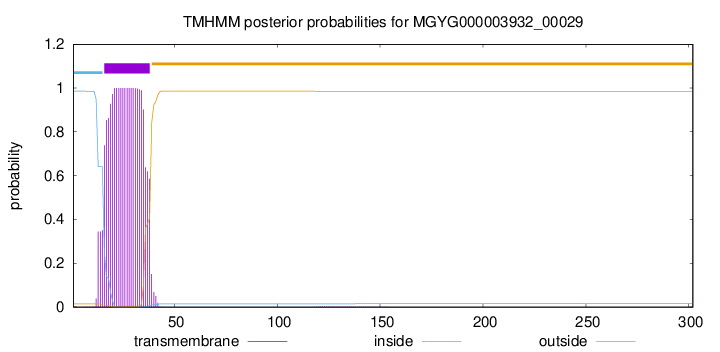
TMHMM annotations
Basic Information help
| Species | CAG-964 sp000435335 | |||||||||||
|---|---|---|---|---|---|---|---|---|---|---|---|---|
| Lineage | Bacteria; Firmicutes_A; Clostridia; Oscillospirales; Acutalibacteraceae; CAG-964; CAG-964 sp000435335 | |||||||||||
| CAZyme ID | MGYG000003932_00029 | |||||||||||
| CAZy Family | CE4 | |||||||||||
| CAZyme Description | Poly-beta-1,6-N-acetyl-D-glucosamine N-deacetylase | |||||||||||
| CAZyme Property |
|
|||||||||||
| Genome Property |
|
|||||||||||
| Gene Location | Start: 29375; End: 30283 Strand: + | |||||||||||
Full Sequence Download help
| MKWVYAMYLN FRVSKYSVVA VVLIITVICF SVIYSGFYPG SSQSTASISS KSDTRGVELP | 60 |
| IVMYHSMLKD TKLQGQFVID PAKFEEDLKY LKDNGYTTIT ASDLIDYVYN KKELPKKPIM | 120 |
| LTFDDGYYNN YLYAYPLLKK YKCKAVISPI VYYSDLYSKS TDAPSPSYSH CTWKQLKEMQ | 180 |
| NSGCVEIQNH SYNMHSQNGR LGIKQKKGES SEEYKEVITQ DISKAQNRFK EKLEYTPQAF | 240 |
| VYPFGALSDS SEEAIKALSF KASFTCEEKI NIIAKDKNSL YLLGRFIRTN KMTTKELFNK | 300 |
| FQ | 302 |
CAZyme Signature Domains help
| Family | Start | End | Evalue | family coverage |
|---|---|---|---|---|
| CE4 | 110 | 263 | 1.6e-26 | 0.9461538461538461 |
CDD Domains download full data without filtering help
| Cdd ID | Domain | E-Value | qStart | qEnd | sStart | sEnd | Domain Description |
|---|---|---|---|---|---|---|---|
| TIGR03938 | deacetyl_PgaB | 2.04e-47 | 64 | 297 | 8 | 257 | poly-beta-1,6-N-acetyl-D-glucosamine N-deacetylase PgaB. Two well-characterized systems produce polysaccharide based on N-acetyl-D-glucosamine in straight chains with beta-1,6 linkages. These are encoded by the icaADBC operon in Staphylococcus species, where the system is designated polysaccharide intercellular adhesin (PIA), and the pgaABCD operon in Gram-negative bacteria such as E. coli. Both systems include a putative polysaccharide deacetylase. The PgaB protein, described here, contains an additional domain lacking from its Gram-positive counterpart IcaB (TIGR03933). Deacetylation by this protein appears necessary to allow export through the porin PgaA [Cell envelope, Biosynthesis and degradation of surface polysaccharides and lipopolysaccharides] |
| cd10918 | CE4_NodB_like_5s_6s | 2.02e-44 | 118 | 286 | 1 | 152 | Putative catalytic NodB homology domain of PgaB, IcaB, and similar proteins which consist of a deformed (beta/alpha)8 barrel fold with 5- or 6-strands. This family belongs to the large and functionally diverse carbohydrate esterase 4 (CE4) superfamily, whose members show strong sequence similarity with some variability due to their distinct carbohydrate substrates. It includes bacterial poly-beta-1,6-N-acetyl-D-glucosamine N-deacetylase PgaB, hemin storage system HmsF protein in gram-negative species, intercellular adhesion proteins IcaB, and many uncharacterized prokaryotic polysaccharide deacetylases. It also includes a putative polysaccharide deacetylase YxkH encoded by the Bacillus subtilis yxkH gene, which is one of six polysaccharide deacetylase gene homologs present in the Bacillus subtilis genome. Sequence comparison shows all family members contain a conserved domain similar to the catalytic NodB homology domain of rhizobial NodB-like proteins, which consists of a deformed (beta/alpha)8 barrel fold with 6 or 7 strands. However, in this family, most proteins have 5 strands and some have 6 strands. Moreover, long insertions are found in many family members, whose function remains unknown. |
| cd10969 | CE4_Ecf1_like_5s | 6.47e-44 | 81 | 286 | 1 | 200 | Putative catalytic NodB homology domain of a hypothetical protein Ecf1 from Escherichia coli and similar proteins. This family contains a hypothetical protein Ecf1 from Escherichia coli and its prokaryotic homologs. Although their biochemical properties remain to be determined, members in this family contain a conserved domain with a 5-stranded beta/alpha barrel, which is similar to the catalytic NodB homology domain of rhizobial NodB-like proteins, belonging to the larger carbohydrate esterase 4 (CE4) superfamily. |
| cd10964 | CE4_PgaB_5s | 7.16e-34 | 114 | 290 | 1 | 192 | N-terminal putative catalytic polysaccharide deacetylase domain of bacterial poly-beta-1,6-N-acetyl-D-glucosamine N-deacetylase PgaB, and similar proteins. This family is represented by an outer membrane lipoprotein, poly-beta-1,6-N-acetyl-D-glucosamine N-deacetylase (PgaB, EC 3.5.1.-), encoded by Escherichia coli pgaB gene from the pgaABCD (formerly ycdSRQP) operon, which affects biofilm development by promoting abiotic surface binding and intercellular adhesion. PgaB catalyzes the N-deacetylation of poly-beta-1,6-N-acetyl-D-glucosamine (PGA), a biofilm adhesin polysaccharide that stabilizes biofilms of E. coli and other bacteria. PgaB contains an N-terminal NodB homology domain with a 5-stranded beta/alpha barrel, and a C-terminal carbohydrate binding domain required for PGA N-deacetylation, which may be involved in binding to unmodified poly-beta-1,6-GlcNAc and assisting catalysis by the deacetylase domain. This family also includes several orthologs of PgaB, such as the hemin storage system HmsF protein, encoded by Yersinia pestis hmsF gene from the hmsHFRS operon, which is essential for Y. pestis biofilm formation. Like PgaB, HmsF is an outer membrane protein with an N-terminal NodB homology domain, which is likely involved in the modification of the exopolysaccharide (EPS) component of the biofilm. HmsF also has a conserved but uncharacterized C-terminal domain that is present in other HmsF-like proteins in Gram-negative bacteria. This alignment model corresponds to the N-terminal NodB homology domain. |
| cd10973 | CE4_DAC_u4_5s | 1.90e-33 | 117 | 286 | 1 | 151 | Putative catalytic NodB homology domain of uncharacterized bacterial polysaccharide deacetylases which consist of a 5-stranded beta/alpha barrel. This family contains many uncharacterized bacterial polysaccharide deacetylases. Although their biological functions remain unknown, all members of the family are predicted to contain a conserved domain with a 5-stranded beta/alpha barrel, which is similar to the catalytic NodB homology domain of rhizobial NodB-like proteins, belonging to the larger carbohydrate esterase 4 (CE4) superfamily. |
CAZyme Hits help
| Hit ID | E-Value | Query Start | Query End | Hit Start | Hit End |
|---|---|---|---|---|---|
| AFS79569.1 | 2.21e-50 | 37 | 300 | 10 | 270 |
| AAM25723.1 | 1.43e-46 | 44 | 271 | 25 | 265 |
| ARI76505.1 | 4.88e-46 | 58 | 284 | 226 | 455 |
| AFS77609.1 | 7.01e-43 | 50 | 297 | 10 | 265 |
| ASF40455.1 | 7.32e-43 | 34 | 300 | 187 | 451 |
PDB Hits download full data without filtering help
| Hit ID | E-Value | Query Start | Query End | Hit Start | Hit End | Description |
|---|---|---|---|---|---|---|
| 4U10_A | 1.45e-25 | 61 | 270 | 6 | 236 | Probingthe structure and mechanism of de-N-acetylase from aggregatibacter actinomycetemcomitans [Aggregatibacter actinomycetemcomitans],4U10_B Probing the structure and mechanism of de-N-acetylase from aggregatibacter actinomycetemcomitans [Aggregatibacter actinomycetemcomitans] |
| 4WCJ_A | 5.25e-21 | 35 | 271 | 12 | 235 | Structureof IcaB from Ammonifex degensii [Ammonifex degensii KC4] |
| 3VUS_A | 2.83e-20 | 62 | 296 | 11 | 259 | Escherichiacoli PgaB N-terminal domain [Escherichia coli K-12],3VUS_B Escherichia coli PgaB N-terminal domain [Escherichia coli K-12] |
| 4F9D_A | 2.50e-19 | 62 | 296 | 15 | 263 | Structureof Escherichia coli PgaB 42-655 in complex with nickel [Escherichia coli K-12],4F9D_B Structure of Escherichia coli PgaB 42-655 in complex with nickel [Escherichia coli K-12] |
| 4F9J_A | 3.37e-19 | 62 | 296 | 15 | 263 | Structureof Escherichia coli PgaB 42-655 in complex with iron [Escherichia coli K-12],4F9J_B Structure of Escherichia coli PgaB 42-655 in complex with iron [Escherichia coli K-12] |
Swiss-Prot Hits download full data without filtering help
| Hit ID | E-Value | Query Start | Query End | Hit Start | Hit End | Description |
|---|---|---|---|---|---|---|
| P75906 | 7.14e-20 | 14 | 296 | 6 | 300 | Poly-beta-1,6-N-acetyl-D-glucosamine N-deacetylase OS=Escherichia coli (strain K12) OX=83333 GN=pgaB PE=1 SV=1 |
| Q8XAR3 | 9.64e-20 | 14 | 296 | 6 | 300 | Poly-beta-1,6-N-acetyl-D-glucosamine N-deacetylase OS=Escherichia coli O157:H7 OX=83334 GN=pgaB PE=3 SV=1 |
| P94361 | 2.08e-16 | 58 | 267 | 64 | 244 | Putative polysaccharide deacetylase YxkH OS=Bacillus subtilis (strain 168) OX=224308 GN=yxkH PE=3 SV=1 |
| P31666 | 5.49e-11 | 59 | 265 | 170 | 370 | Uncharacterized protein YadE OS=Escherichia coli (strain K12) OX=83333 GN=yadE PE=3 SV=2 |
SignalP and Lipop Annotations help
This protein is predicted as OTHER
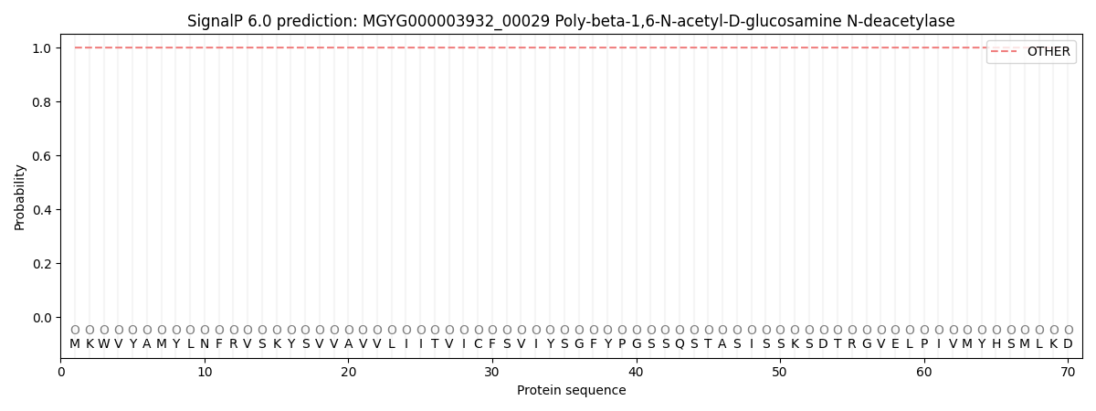
| Other | SP_Sec_SPI | LIPO_Sec_SPII | TAT_Tat_SPI | TATLIP_Sec_SPII | PILIN_Sec_SPIII |
|---|---|---|---|---|---|
| 1.000011 | 0.000009 | 0.000000 | 0.000000 | 0.000000 | 0.000001 |