You are browsing environment: HUMAN GUT
CAZyme Information: MGYG000002956_00167
You are here: Home > Sequence: MGYG000002956_00167
Basic Information |
Genomic context |
Full Sequence |
Enzyme annotations |
CAZy signature domains |
CDD domains |
CAZyme hits |
PDB hits |
Swiss-Prot hits |
SignalP and Lipop annotations |
TMHMM annotations
Basic Information help
| Species | Lactobacillus kalixensis | |||||||||||
|---|---|---|---|---|---|---|---|---|---|---|---|---|
| Lineage | Bacteria; Firmicutes; Bacilli; Lactobacillales; Lactobacillaceae; Lactobacillus; Lactobacillus kalixensis | |||||||||||
| CAZyme ID | MGYG000002956_00167 | |||||||||||
| CAZy Family | GH1 | |||||||||||
| CAZyme Description | Aryl-phospho-beta-D-glucosidase BglH | |||||||||||
| CAZyme Property |
|
|||||||||||
| Genome Property |
|
|||||||||||
| Gene Location | Start: 3193; End: 4665 Strand: - | |||||||||||
Full Sequence Download help
| MTFPKNFLWG GATAANQIEG AYDVDGKGLS VTDIMTAGGL DHPRLLTYKM DGKLQKSPSA | 60 |
| PGQGLPEGAV GAIDHNEYYP NHVAIDFYHH YKEDIKMFAD MGFKTFRLSI AWTRIFPNGD | 120 |
| DEQPNQVGLD FYRHVFEECK KYDIEPLVTI SHYEDPLHLS EKYNDWQDRG MIDCYVKYAK | 180 |
| TLFKEYKGLV KYWLTFNEIN SAVLFLNMFG GNGTDEDYQH AYQKLHYQFV ASARAVKLAH | 240 |
| EIDPNYVIGN MICGIVDYPM TPDPKDVLLN RHDWEQDLFY CGDAQCKGKY PTFAKRLWDE | 300 |
| HNVHLDITEQ DEKDMLEGKV DMYTFSYYMS NVVTTHEVKD KVGGNFAAGA KNPYLKYSEW | 360 |
| GWATDPTGLQ YYLEVMYDRY ELPIMIVENG LGAVDKISDD GKIHDDYRID YLRLHIEAMQ | 420 |
| KAIDDGVDLI GYTTWGCIDC VSAGTGQMSK RYGFIYVDRD DEGNGSLDRM PKDSFYWYKK | 480 |
| VIESNGNDLD | 490 |
CAZyme Signature Domains help
| Family | Start | End | Evalue | family coverage |
|---|---|---|---|---|
| GH1 | 2 | 486 | 6.8e-145 | 0.9883449883449883 |
CDD Domains download full data without filtering help
| Cdd ID | Domain | E-Value | qStart | qEnd | sStart | sEnd | Domain Description |
|---|---|---|---|---|---|---|---|
| PRK09589 | celA | 0.0 | 1 | 489 | 2 | 476 | 6-phospho-beta-glucosidase; Reviewed |
| PRK09852 | PRK09852 | 0.0 | 2 | 490 | 3 | 474 | cryptic 6-phospho-beta-glucosidase; Provisional |
| COG2723 | BglB | 0.0 | 1 | 490 | 2 | 458 | Beta-glucosidase/6-phospho-beta-glucosidase/beta-galactosidase [Carbohydrate transport and metabolism]. |
| PRK09593 | arb | 0.0 | 1 | 490 | 4 | 478 | 6-phospho-beta-glucosidase; Reviewed |
| PRK15014 | PRK15014 | 5.64e-174 | 1 | 489 | 4 | 477 | 6-phospho-beta-glucosidase BglA; Provisional |
CAZyme Hits help
| Hit ID | E-Value | Query Start | Query End | Hit Start | Hit End |
|---|---|---|---|---|---|
| QXL47430.1 | 0.0 | 1 | 490 | 1 | 490 |
| CAX67457.1 | 0.0 | 1 | 490 | 1 | 490 |
| QGY97222.1 | 0.0 | 1 | 490 | 1 | 490 |
| QGT98326.1 | 0.0 | 1 | 490 | 1 | 490 |
| ARW74425.1 | 0.0 | 1 | 490 | 1 | 490 |
PDB Hits download full data without filtering help
| Hit ID | E-Value | Query Start | Query End | Hit Start | Hit End | Description |
|---|---|---|---|---|---|---|
| 6WGD_A | 3.01e-165 | 3 | 489 | 8 | 469 | Crystalstructure of a 6-phospho-beta-glucosidase from Bacillus licheniformis [Bacillus licheniformis],6WGD_B Crystal structure of a 6-phospho-beta-glucosidase from Bacillus licheniformis [Bacillus licheniformis],6WGD_C Crystal structure of a 6-phospho-beta-glucosidase from Bacillus licheniformis [Bacillus licheniformis] |
| 4F66_A | 2.32e-162 | 4 | 489 | 8 | 480 | Thecrystal structure of 6-phospho-beta-glucosidase from Streptococcus mutans UA159 in complex with beta-D-glucose-6-phosphate. [Streptococcus mutans],4F66_B The crystal structure of 6-phospho-beta-glucosidase from Streptococcus mutans UA159 in complex with beta-D-glucose-6-phosphate. [Streptococcus mutans] |
| 4F79_A | 6.60e-162 | 4 | 489 | 8 | 480 | Thecrystal structure of 6-phospho-beta-glucosidase mutant (E375Q) in complex with Salicin 6-phosphate [Streptococcus mutans],4GPN_A The crystal structure of 6-P-beta-D-Glucosidase (E375Q mutant) from Streptococcus mutans UA150 in complex with Gentiobiose 6-phosphate. [Streptococcus mutans UA159],4GPN_B The crystal structure of 6-P-beta-D-Glucosidase (E375Q mutant) from Streptococcus mutans UA150 in complex with Gentiobiose 6-phosphate. [Streptococcus mutans UA159] |
| 3PN8_A | 5.24e-156 | 4 | 489 | 8 | 480 | Thecrystal structure of 6-phospho-beta-glucosidase from Streptococcus mutans UA159 [Streptococcus mutans],3PN8_B The crystal structure of 6-phospho-beta-glucosidase from Streptococcus mutans UA159 [Streptococcus mutans] |
| 2XHY_A | 1.16e-154 | 1 | 489 | 6 | 479 | CrystalStructure of E.coli BglA [Escherichia coli K-12],2XHY_B Crystal Structure of E.coli BglA [Escherichia coli K-12],2XHY_C Crystal Structure of E.coli BglA [Escherichia coli K-12],2XHY_D Crystal Structure of E.coli BglA [Escherichia coli K-12] |
Swiss-Prot Hits download full data without filtering help
| Hit ID | E-Value | Query Start | Query End | Hit Start | Hit End | Description |
|---|---|---|---|---|---|---|
| Q46130 | 1.09e-168 | 1 | 490 | 5 | 472 | 6-phospho-beta-glucosidase OS=Clostridium longisporum OX=1523 GN=abgA PE=3 SV=1 |
| P40740 | 4.63e-159 | 3 | 489 | 8 | 469 | Aryl-phospho-beta-D-glucosidase BglH OS=Bacillus subtilis (strain 168) OX=224308 GN=bglH PE=1 SV=2 |
| P26206 | 2.81e-154 | 3 | 489 | 5 | 463 | 6-phospho-beta-glucosidase OS=Dickeya chrysanthemi OX=556 GN=arbB PE=3 SV=1 |
| Q46829 | 6.37e-154 | 1 | 489 | 6 | 479 | 6-phospho-beta-glucosidase BglA OS=Escherichia coli (strain K12) OX=83333 GN=bglA PE=1 SV=2 |
| P42973 | 1.03e-152 | 4 | 489 | 5 | 479 | Aryl-phospho-beta-D-glucosidase BglA OS=Bacillus subtilis (strain 168) OX=224308 GN=bglA PE=1 SV=1 |
SignalP and Lipop Annotations help
This protein is predicted as OTHER
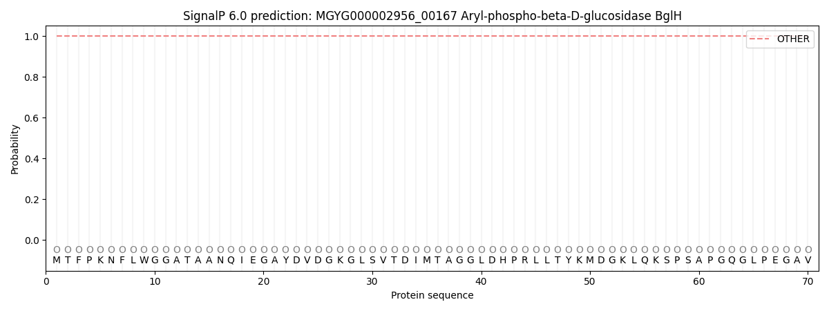
| Other | SP_Sec_SPI | LIPO_Sec_SPII | TAT_Tat_SPI | TATLIP_Sec_SPII | PILIN_Sec_SPIII |
|---|---|---|---|---|---|
| 1.000059 | 0.000000 | 0.000000 | 0.000000 | 0.000000 | 0.000000 |