You are browsing environment: HUMAN GUT
CAZyme Information: MGYG000000091_00402
You are here: Home > Sequence: MGYG000000091_00402
Basic Information |
Genomic context |
Full Sequence |
Enzyme annotations |
CAZy signature domains |
CDD domains |
CAZyme hits |
PDB hits |
Swiss-Prot hits |
SignalP and Lipop annotations |
TMHMM annotations
Basic Information help
| Species | Achromobacter xylosoxidans | |||||||||||
|---|---|---|---|---|---|---|---|---|---|---|---|---|
| Lineage | Bacteria; Proteobacteria; Gammaproteobacteria; Burkholderiales; Burkholderiaceae; Achromobacter; Achromobacter xylosoxidans | |||||||||||
| CAZyme ID | MGYG000000091_00402 | |||||||||||
| CAZy Family | GT0 | |||||||||||
| CAZyme Description | UDP-N-acetylglucosamine 2-epimerase | |||||||||||
| CAZyme Property |
|
|||||||||||
| Genome Property |
|
|||||||||||
| Gene Location | Start: 472071; End: 473186 Strand: - | |||||||||||
Full Sequence Download help
| MAPLVAALQE ESRIDSVVCV TGQHRQMLDQ VLALFDLRAR HDLDIMVPNQ TLNGLYARLI | 60 |
| SQVDGVLEKE QPDYVLVHGD TSTASACALA AFHRRIRIGH VEAGLRTGNL AMPFPEEMNR | 120 |
| RVVDAIGDAL FAPTAESRAN LLRENLAGRI TVTGNTVIDA LALTCAKLAP DGPLARGLEA | 180 |
| RYHWLDARRR LMLVTGHRRE SFGGGFKNIC AALAELARRE DLQIVYPVHL NPQVRNVVMK | 240 |
| DLAGLERVHL IDPLDYLDFV WFMQRAYLIL TDSGGVQEEA PYLGKPVLVM RDVTERPEAV | 300 |
| QAGTVALVGT DTQRILTEVN RLLDDPALHA SFSRRINPYG DGKASGRIVD ALCGRPVSEF | 360 |
| DPHGPAAGTA A | 371 |
CDD Domains download full data without filtering help
| Cdd ID | Domain | E-Value | qStart | qEnd | sStart | sEnd | Domain Description |
|---|---|---|---|---|---|---|---|
| COG0381 | WecB | 3.46e-156 | 1 | 352 | 19 | 368 | UDP-N-acetylglucosamine 2-epimerase [Cell wall/membrane/envelope biogenesis]. |
| pfam02350 | Epimerase_2 | 1.85e-151 | 7 | 352 | 2 | 335 | UDP-N-acetylglucosamine 2-epimerase. This family consists of UDP-N-acetylglucosamine 2-epimerases EC:5.1.3.14 this enzyme catalyzes the production of UDP-ManNAc from UDP-GlcNAc. Note that some of the enzymes is this family are bifunctional, in these instances Pfam matches only the N-terminal half of the protein suggesting that the additional C-terminal part (when compared to mono-functional members of this family) is responsible for the UPD-N-acetylmannosamine kinase activity of these enzymes. This hypothesis is further supported by the assumption that the C-terminal part of rat Gne is the kinase domain. |
| TIGR00236 | wecB | 9.12e-148 | 1 | 352 | 16 | 361 | UDP-N-acetylglucosamine 2-epimerase. This cytosolic enzyme converts UDP-N-acetyl-D-glucosamine to UDP-N-acetyl-D-mannosamine. In E. coli, this is the first step in the pathway of enterobacterial common antigen biosynthesis.Members of this orthology group have many gene symbols, often reflecting the overall activity of the pathway and/or operon that includes it. Symbols include epsC (exopolysaccharide C) in Burkholderia solanacerum, cap8P (type 8 capsule P) in Staphylococcus aureus, and nfrC in an older designation based on the effects of deletion on phage N4 adsorption. Epimerase activity was also demonstrated in a bifunctional rat enzyme, for which the N-terminal domain appears to be orthologous. The set of proteins found above the suggested cutoff includes E. coli WecB in one of two deeply branched clusters and the rat UDP-N-acetylglucosamine 2-epimerase domain in the other. [Cell envelope, Biosynthesis and degradation of surface polysaccharides and lipopolysaccharides] |
| cd03786 | GTB_UDP-GlcNAc_2-Epimerase | 1.81e-146 | 1 | 352 | 15 | 364 | UDP-N-acetylglucosamine 2-epimerase and similar proteins. Bacterial members of the UDP-N-Acetylglucosamine (GlcNAc) 2-Epimerase family (EC 5.1.3.14) are known to catalyze the reversible interconversion of UDP-GlcNAc and UDP-N-acetylmannosamine (UDP-ManNAc). The enzyme serves to produce an activated form of ManNAc residues (UDP-ManNAc) for use in the biosynthesis of a variety of cell surface polysaccharides; The mammalian enzyme is bifunctional, catalyzing both the inversion of stereochemistry at C-2 and the hydrolysis of the UDP-sugar linkage to generate free ManNAc. It also catalyzes the phosphorylation of ManNAc to generate ManNAc 6-phosphate, a precursor to salic acids. In mammals, sialic acids are found at the termini of oligosaccharides in a large variety of cell surface glycoconjugates and are key mediators of cell-cell recognition events. Mutations in human members of this family have been associated with Sialuria, a rare disease caused by the disorders of sialic acid metabolism. This family belongs to the GT-B structural superfamily of glycoslytransferases, which have characteristic N- and C-terminal domains each containing a typical Rossmann fold. The two domains have high structural homology despite minimal sequence homology. The large cleft that separates the two domains includes the catalytic center and permits a high degree of flexibility. |
| cd03801 | GT4_PimA-like | 0.004 | 38 | 335 | 49 | 343 | phosphatidyl-myo-inositol mannosyltransferase. This family is most closely related to the GT4 family of glycosyltransferases and named after PimA in Propionibacterium freudenreichii, which is involved in the biosynthesis of phosphatidyl-myo-inositol mannosides (PIM) which are early precursors in the biosynthesis of lipomannans (LM) and lipoarabinomannans (LAM), and catalyzes the addition of a mannosyl residue from GDP-D-mannose (GDP-Man) to the position 2 of the carrier lipid phosphatidyl-myo-inositol (PI) to generate a phosphatidyl-myo-inositol bearing an alpha-1,2-linked mannose residue (PIM1). Glycosyltransferases catalyze the transfer of sugar moieties from activated donor molecules to specific acceptor molecules, forming glycosidic bonds. The acceptor molecule can be a lipid, a protein, a heterocyclic compound, or another carbohydrate residue. This group of glycosyltransferases is most closely related to the previously defined glycosyltransferase family 1 (GT1). The members of this family may transfer UDP, ADP, GDP, or CMP linked sugars. The diverse enzymatic activities among members of this family reflect a wide range of biological functions. The protein structure available for this family has the GTB topology, one of the two protein topologies observed for nucleotide-sugar-dependent glycosyltransferases. GTB proteins have distinct N- and C- terminal domains each containing a typical Rossmann fold. The two domains have high structural homology despite minimal sequence homology. The large cleft that separates the two domains includes the catalytic center and permits a high degree of flexibility. The members of this family are found mainly in certain bacteria and archaea. |
CAZyme Hits help
| Hit ID | E-Value | Query Start | Query End | Hit Start | Hit End |
|---|---|---|---|---|---|
| QLK59446.1 | 2.45e-134 | 1 | 359 | 16 | 376 |
| QUT15423.1 | 4.92e-134 | 1 | 359 | 16 | 376 |
| QQN33861.1 | 3.24e-132 | 1 | 359 | 16 | 376 |
| AZP48621.1 | 4.60e-132 | 1 | 359 | 16 | 376 |
| AZP44285.1 | 4.60e-132 | 1 | 359 | 16 | 376 |
PDB Hits download full data without filtering help
| Hit ID | E-Value | Query Start | Query End | Hit Start | Hit End | Description |
|---|---|---|---|---|---|---|
| 3DZC_A | 2.17e-131 | 1 | 352 | 41 | 394 | 2.35Angstrom resolution structure of WecB (VC0917), a UDP-N-acetylglucosamine 2-epimerase from Vibrio cholerae. [Vibrio cholerae],3DZC_B 2.35 Angstrom resolution structure of WecB (VC0917), a UDP-N-acetylglucosamine 2-epimerase from Vibrio cholerae. [Vibrio cholerae] |
| 5DLD_A | 6.60e-127 | 1 | 370 | 25 | 396 | CrystalStructure of a UDP-N-acetylglucosamine 2-epimerase from Burkholderia vietnamiensis complexed with UDP-GlcNAc and UDP [Burkholderia vietnamiensis G4] |
| 1F6D_A | 1.80e-125 | 2 | 358 | 17 | 375 | TheStructure Of Udp-N-Acetylglucosamine 2-Epimerase From E. Coli. [Escherichia coli],1F6D_B The Structure Of Udp-N-Acetylglucosamine 2-Epimerase From E. Coli. [Escherichia coli],1F6D_C The Structure Of Udp-N-Acetylglucosamine 2-Epimerase From E. Coli. [Escherichia coli],1F6D_D The Structure Of Udp-N-Acetylglucosamine 2-Epimerase From E. Coli. [Escherichia coli] |
| 1VGV_A | 2.35e-125 | 2 | 358 | 17 | 375 | Crystalstructure of UDP-N-acetylglucosamine_2 epimerase [Escherichia coli],1VGV_B Crystal structure of UDP-N-acetylglucosamine_2 epimerase [Escherichia coli],1VGV_C Crystal structure of UDP-N-acetylglucosamine_2 epimerase [Escherichia coli],1VGV_D Crystal structure of UDP-N-acetylglucosamine_2 epimerase [Escherichia coli] |
| 6VLB_A | 3.23e-114 | 1 | 352 | 16 | 368 | Crystalstructure of ligand-free UDP-GlcNAc 2-epimerase from Neisseria meningitidis [Neisseria meningitidis Z2491],6VLB_B Crystal structure of ligand-free UDP-GlcNAc 2-epimerase from Neisseria meningitidis [Neisseria meningitidis Z2491],6VLC_A Crystal structure of UDP-GlcNAc 2-epimerase from Neisseria meningitidis bound to UDP-GlcNAc [Neisseria meningitidis Z2491],6VLC_B Crystal structure of UDP-GlcNAc 2-epimerase from Neisseria meningitidis bound to UDP-GlcNAc [Neisseria meningitidis Z2491],6VLC_C Crystal structure of UDP-GlcNAc 2-epimerase from Neisseria meningitidis bound to UDP-GlcNAc [Neisseria meningitidis Z2491],6VLC_D Crystal structure of UDP-GlcNAc 2-epimerase from Neisseria meningitidis bound to UDP-GlcNAc [Neisseria meningitidis Z2491] |
Swiss-Prot Hits download full data without filtering help
| Hit ID | E-Value | Query Start | Query End | Hit Start | Hit End | Description |
|---|---|---|---|---|---|---|
| P58600 | 2.37e-130 | 1 | 352 | 17 | 370 | Probable UDP-N-acetylglucosamine 2-epimerase OS=Ralstonia solanacearum (strain GMI1000) OX=267608 GN=epsC PE=3 SV=1 |
| Q8XAR8 | 1.98e-129 | 1 | 358 | 16 | 375 | UDP-N-acetylglucosamine 2-epimerase OS=Escherichia coli O157:H7 OX=83334 GN=wecB PE=3 SV=1 |
| P27828 | 1.98e-129 | 1 | 358 | 16 | 375 | UDP-N-acetylglucosamine 2-epimerase OS=Escherichia coli (strain K12) OX=83333 GN=wecB PE=1 SV=2 |
| P52641 | 2.72e-129 | 1 | 352 | 17 | 370 | Probable UDP-N-acetylglucosamine 2-epimerase OS=Ralstonia solanacearum OX=305 GN=epsC PE=3 SV=2 |
| Q8ZAE3 | 8.01e-129 | 1 | 358 | 16 | 375 | UDP-N-acetylglucosamine 2-epimerase OS=Yersinia pestis OX=632 GN=wecB PE=3 SV=1 |
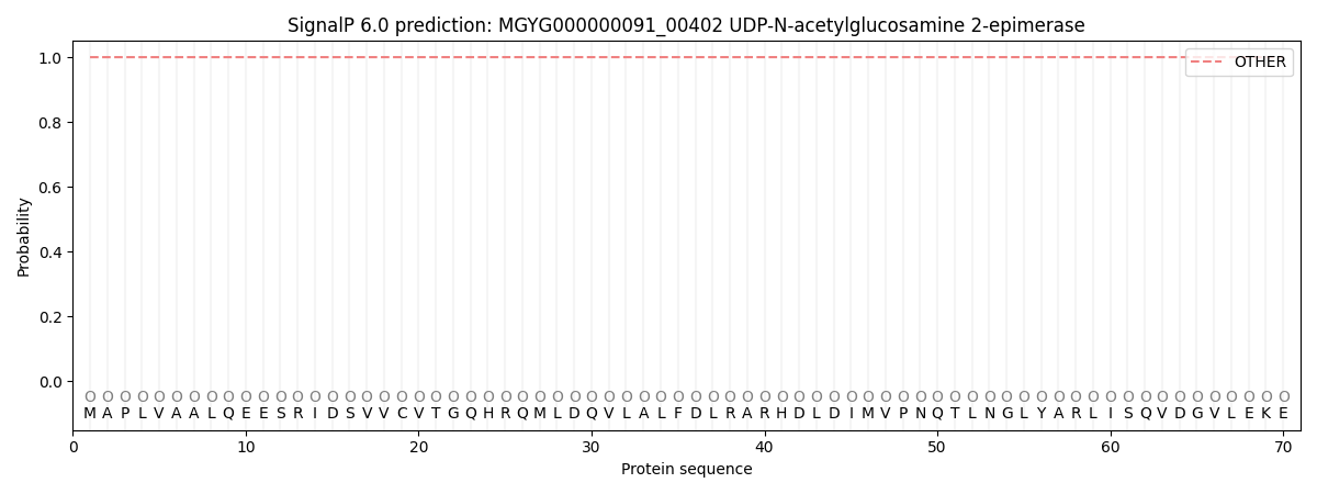
SignalP and Lipop Annotations help
This protein is predicted as OTHER
| Other | SP_Sec_SPI | LIPO_Sec_SPII | TAT_Tat_SPI | TATLIP_Sec_SPII | PILIN_Sec_SPIII |
|---|---|---|---|---|---|
| 1.000064 | 0.000000 | 0.000000 | 0.000000 | 0.000000 | 0.000000 |