You are browsing environment: HUMAN GUT
CAZyme Information: MGYG000003659_00145
You are here: Home > Sequence: MGYG000003659_00145
Basic Information |
Genomic context |
Full Sequence |
Enzyme annotations |
CAZy signature domains |
CDD domains |
CAZyme hits |
PDB hits |
Swiss-Prot hits |
SignalP and Lipop annotations |
TMHMM annotations
Basic Information help
| Species | CAG-594 sp900771805 | |||||||||||
|---|---|---|---|---|---|---|---|---|---|---|---|---|
| Lineage | Bacteria; Firmicutes; Bacilli; RF39; UBA660; CAG-594; CAG-594 sp900771805 | |||||||||||
| CAZyme ID | MGYG000003659_00145 | |||||||||||
| CAZy Family | GT0 | |||||||||||
| CAZyme Description | UDP-N-acetylglucosamine 2-epimerase | |||||||||||
| CAZyme Property |
|
|||||||||||
| Genome Property |
|
|||||||||||
| Gene Location | Start: 75759; End: 76868 Strand: + | |||||||||||
Full Sequence Download help
| MIKVMSIFGT RPEAIKMAPL VKELESRKEI ESIVCVTAQH REMLDQVLET FDIKPDYDLN | 60 |
| IMKQGQTLAE VTTRALLGLE EVIKEVKPDI VLVHGDTTTT FAGALAAFYN QVAIGHVEAG | 120 |
| LRTYDKYSPF PEEMNRQMVD CMTDMYFAPT NLSKSNLLKQ NIEESKIYVT GNTAIDAMKT | 180 |
| TVDKDYSNEV LDWVGNDRMI LLTAHRRENL GDPMRHIFKA IKRIVDEFDD IKVVYPIHMN | 240 |
| PRVREVAKEI FSNCDRIRLI EPLEVFDFHN FQNKSYIILS DSGGIQEEAP SLGKPVLVLR | 300 |
| DTTERPEGIE AGTLKLVGTN EETIYKETKK LLTDKAEYDR MSKASNPYGD GHTSERIADA | 360 |
| IIEKFNKEY | 369 |
CDD Domains download full data without filtering help
| Cdd ID | Domain | E-Value | qStart | qEnd | sStart | sEnd | Domain Description |
|---|---|---|---|---|---|---|---|
| COG0381 | WecB | 0.0 | 1 | 366 | 3 | 373 | UDP-N-acetylglucosamine 2-epimerase [Cell wall/membrane/envelope biogenesis]. |
| TIGR00236 | wecB | 0.0 | 2 | 361 | 1 | 361 | UDP-N-acetylglucosamine 2-epimerase. This cytosolic enzyme converts UDP-N-acetyl-D-glucosamine to UDP-N-acetyl-D-mannosamine. In E. coli, this is the first step in the pathway of enterobacterial common antigen biosynthesis.Members of this orthology group have many gene symbols, often reflecting the overall activity of the pathway and/or operon that includes it. Symbols include epsC (exopolysaccharide C) in Burkholderia solanacerum, cap8P (type 8 capsule P) in Staphylococcus aureus, and nfrC in an older designation based on the effects of deletion on phage N4 adsorption. Epimerase activity was also demonstrated in a bifunctional rat enzyme, for which the N-terminal domain appears to be orthologous. The set of proteins found above the suggested cutoff includes E. coli WecB in one of two deeply branched clusters and the rat UDP-N-acetylglucosamine 2-epimerase domain in the other. [Cell envelope, Biosynthesis and degradation of surface polysaccharides and lipopolysaccharides] |
| cd03786 | GTB_UDP-GlcNAc_2-Epimerase | 1.87e-168 | 3 | 362 | 1 | 365 | UDP-N-acetylglucosamine 2-epimerase and similar proteins. Bacterial members of the UDP-N-Acetylglucosamine (GlcNAc) 2-Epimerase family (EC 5.1.3.14) are known to catalyze the reversible interconversion of UDP-GlcNAc and UDP-N-acetylmannosamine (UDP-ManNAc). The enzyme serves to produce an activated form of ManNAc residues (UDP-ManNAc) for use in the biosynthesis of a variety of cell surface polysaccharides; The mammalian enzyme is bifunctional, catalyzing both the inversion of stereochemistry at C-2 and the hydrolysis of the UDP-sugar linkage to generate free ManNAc. It also catalyzes the phosphorylation of ManNAc to generate ManNAc 6-phosphate, a precursor to salic acids. In mammals, sialic acids are found at the termini of oligosaccharides in a large variety of cell surface glycoconjugates and are key mediators of cell-cell recognition events. Mutations in human members of this family have been associated with Sialuria, a rare disease caused by the disorders of sialic acid metabolism. This family belongs to the GT-B structural superfamily of glycoslytransferases, which have characteristic N- and C-terminal domains each containing a typical Rossmann fold. The two domains have high structural homology despite minimal sequence homology. The large cleft that separates the two domains includes the catalytic center and permits a high degree of flexibility. |
| pfam02350 | Epimerase_2 | 1.55e-159 | 22 | 361 | 1 | 335 | UDP-N-acetylglucosamine 2-epimerase. This family consists of UDP-N-acetylglucosamine 2-epimerases EC:5.1.3.14 this enzyme catalyzes the production of UDP-ManNAc from UDP-GlcNAc. Note that some of the enzymes is this family are bifunctional, in these instances Pfam matches only the N-terminal half of the protein suggesting that the additional C-terminal part (when compared to mono-functional members of this family) is responsible for the UPD-N-acetylmannosamine kinase activity of these enzymes. This hypothesis is further supported by the assumption that the C-terminal part of rat Gne is the kinase domain. |
| cd17507 | GT28_Beta-DGS-like | 3.41e-05 | 79 | 363 | 89 | 361 | beta-diglucosyldiacylglycerol synthase and similar proteins. beta-diglucosyldiacylglycerol synthase (processive diacylglycerol beta-glucosyltransferase EC 2.4.1.315) is involved in the biosynthesis of both the bilayer- and non-bilayer-forming membrane glucolipids. This family of glycosyltransferases also contains plant major galactolipid synthase (chloroplastic monogalactosyldiacylglycerol synthase 1 EC 2.4.1.46). Glycosyltransferases catalyze the transfer of sugar moieties from activated donor molecules to specific acceptor molecules, forming glycosidic bonds. The acceptor molecule can be a lipid, a protein, a heterocyclic compound, or another carbohydrate residue. The structures of the formed glycoconjugates are extremely diverse, reflecting a wide range of biological functions. The members of this family share a common GTB topology, one of the two protein topologies observed for nucleotide-sugar-dependent glycosyltransferases. GTB proteins have distinct N- and C- terminal domains each containing a typical Rossmann fold. The two domains have high structural homology despite minimal sequence homology. The large cleft that separates the two domains includes the catalytic center and permits a high degree of flexibility. |
CAZyme Hits help
| Hit ID | E-Value | Query Start | Query End | Hit Start | Hit End |
|---|---|---|---|---|---|
| QLA08260.1 | 6.58e-177 | 3 | 361 | 3 | 361 |
| AUV68874.1 | 1.26e-175 | 3 | 361 | 3 | 361 |
| AUV66492.1 | 1.26e-175 | 3 | 361 | 3 | 361 |
| AMW24368.1 | 7.24e-175 | 3 | 361 | 3 | 361 |
| AVH46190.1 | 2.94e-174 | 3 | 361 | 3 | 361 |
PDB Hits download full data without filtering help
| Hit ID | E-Value | Query Start | Query End | Hit Start | Hit End | Description |
|---|---|---|---|---|---|---|
| 4FKZ_A | 7.44e-183 | 2 | 362 | 4 | 364 | Crystalstructure of Bacillus subtilis UDP-GlcNAc 2-epimerase in complex with UDP-GlcNAc and UDP [Bacillus subtilis subsp. subtilis str. 168],4FKZ_B Crystal structure of Bacillus subtilis UDP-GlcNAc 2-epimerase in complex with UDP-GlcNAc and UDP [Bacillus subtilis subsp. subtilis str. 168] |
| 3BEO_A | 2.69e-182 | 2 | 367 | 9 | 375 | AStructural Basis for the allosteric regulation of non-hydrolyzing UDP-GlcNAc 2-epimerases [Bacillus anthracis],3BEO_B A Structural Basis for the allosteric regulation of non-hydrolyzing UDP-GlcNAc 2-epimerases [Bacillus anthracis] |
| 1O6C_A | 2.98e-175 | 2 | 362 | 4 | 364 | Crystalstructure of UDP-N-acetylglucosamine 2-epimerase [Bacillus subtilis],1O6C_B Crystal structure of UDP-N-acetylglucosamine 2-epimerase [Bacillus subtilis] |
| 5ENZ_A | 2.77e-170 | 3 | 361 | 3 | 361 | S.aureus MnaA-UDP co-structure [Staphylococcus aureus],5ENZ_B S. aureus MnaA-UDP co-structure [Staphylococcus aureus] |
| 3OT5_A | 1.08e-164 | 2 | 367 | 28 | 394 | 2.2Angstrom Resolution Crystal Structure of putative UDP-N-acetylglucosamine 2-epimerase from Listeria monocytogenes [Listeria monocytogenes EGD-e],3OT5_B 2.2 Angstrom Resolution Crystal Structure of putative UDP-N-acetylglucosamine 2-epimerase from Listeria monocytogenes [Listeria monocytogenes EGD-e],3OT5_C 2.2 Angstrom Resolution Crystal Structure of putative UDP-N-acetylglucosamine 2-epimerase from Listeria monocytogenes [Listeria monocytogenes EGD-e],3OT5_D 2.2 Angstrom Resolution Crystal Structure of putative UDP-N-acetylglucosamine 2-epimerase from Listeria monocytogenes [Listeria monocytogenes EGD-e] |
Swiss-Prot Hits download full data without filtering help
| Hit ID | E-Value | Query Start | Query End | Hit Start | Hit End | Description |
|---|---|---|---|---|---|---|
| P39131 | 3.06e-182 | 2 | 362 | 4 | 364 | UDP-N-acetylglucosamine 2-epimerase OS=Bacillus subtilis (strain 168) OX=224308 GN=mnaA PE=1 SV=1 |
| Q9X0C4 | 5.93e-158 | 1 | 365 | 1 | 368 | Putative UDP-N-acetylglucosamine 2-epimerase OS=Thermotoga maritima (strain ATCC 43589 / DSM 3109 / JCM 10099 / NBRC 100826 / MSB8) OX=243274 GN=TM_1034 PE=3 SV=1 |
| P45360 | 1.70e-149 | 2 | 365 | 4 | 371 | Putative UDP-N-acetylglucosamine 2-epimerase OS=Clostridium acetobutylicum (strain ATCC 824 / DSM 792 / JCM 1419 / LMG 5710 / VKM B-1787) OX=272562 GN=CA_C2874 PE=3 SV=2 |
| Q8XAR8 | 5.63e-131 | 3 | 361 | 2 | 369 | UDP-N-acetylglucosamine 2-epimerase OS=Escherichia coli O157:H7 OX=83334 GN=wecB PE=3 SV=1 |
| P27828 | 1.13e-130 | 3 | 361 | 2 | 369 | UDP-N-acetylglucosamine 2-epimerase OS=Escherichia coli (strain K12) OX=83333 GN=wecB PE=1 SV=2 |
SignalP and Lipop Annotations help
This protein is predicted as OTHER
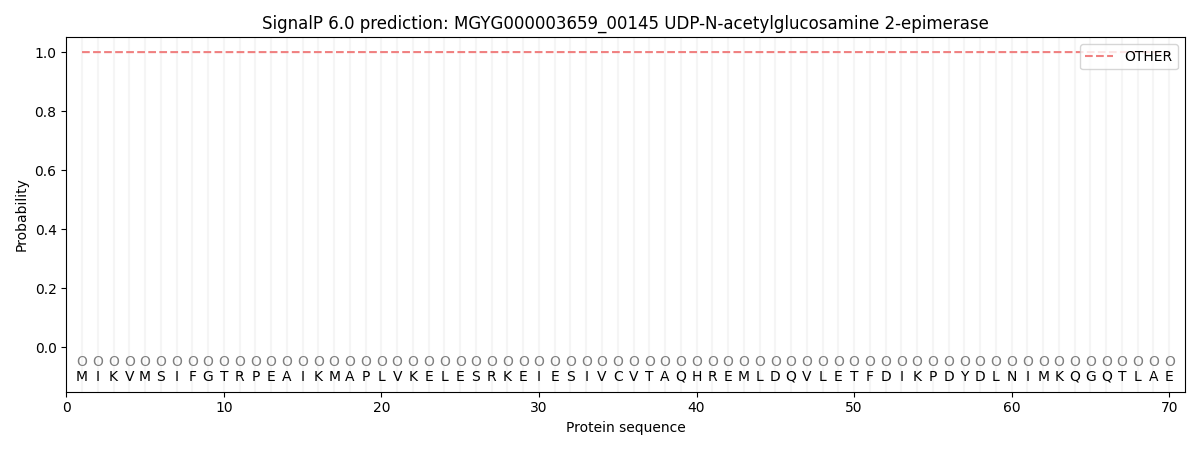
| Other | SP_Sec_SPI | LIPO_Sec_SPII | TAT_Tat_SPI | TATLIP_Sec_SPII | PILIN_Sec_SPIII |
|---|---|---|---|---|---|
| 1.000035 | 0.000000 | 0.000000 | 0.000000 | 0.000000 | 0.000000 |