You are browsing environment: HUMAN GUT
CAZyme Information: MGYG000004714_00831
You are here: Home > Sequence: MGYG000004714_00831
Basic Information |
Genomic context |
Full Sequence |
Enzyme annotations |
CAZy signature domains |
CDD domains |
CAZyme hits |
PDB hits |
Swiss-Prot hits |
SignalP and Lipop annotations |
TMHMM annotations
Basic Information help
| Species | ||||||||||||
|---|---|---|---|---|---|---|---|---|---|---|---|---|
| Lineage | Bacteria; Firmicutes_A; Clostridia; Clostridiales; Clostridiaceae; Clostridium_X; | |||||||||||
| CAZyme ID | MGYG000004714_00831 | |||||||||||
| CAZy Family | GT0 | |||||||||||
| CAZyme Description | UDP-N-acetylglucosamine 2-epimerase | |||||||||||
| CAZyme Property |
|
|||||||||||
| Genome Property |
|
|||||||||||
| Gene Location | Start: 7; End: 1194 Strand: - | |||||||||||
Full Sequence Download help
| MENGIFQEKY LALGEKIMKK IKVMTIFGTR PEAIKMAPLV KELESRDEIE SIVCVTAQHR | 60 |
| QMLDQVLNLF EITPQYDLNI MQTKQTLTGI TNRVLEGLEK IYDDVEPDMI LVHGDTTTTF | 120 |
| AGALSAFYKQ IKVGHVEAGL RTFDKYFPFP EEMNRKLTGA IADLHFAPTQ GSKANLLREG | 180 |
| VGEDQVIITG NTVIDAMEFT VEDDYKFETE ELNTIDFENK KVIMVTAHRR ENWGEGIENI | 240 |
| CKSLKSIVEN NKDVELNPVV KDVVYKHLDN MNRVHLLSPL DTKETHNLMN KCFMVMTDSG | 300 |
| GLQEEAPHLG KPVLVLRDVT ERPEAVEAGT VKLLGTDFQR ICEEANKLLN DEEAYKAMSK | 360 |
| AINPYGDGLA SKRIVDSILK YYKITDRIIE EFKIK | 395 |
CDD Domains download full data without filtering help
| Cdd ID | Domain | E-Value | qStart | qEnd | sStart | sEnd | Domain Description |
|---|---|---|---|---|---|---|---|
| COG0381 | WecB | 0.0 | 18 | 393 | 1 | 383 | UDP-N-acetylglucosamine 2-epimerase [Cell wall/membrane/envelope biogenesis]. |
| TIGR00236 | wecB | 4.13e-173 | 21 | 382 | 1 | 365 | UDP-N-acetylglucosamine 2-epimerase. This cytosolic enzyme converts UDP-N-acetyl-D-glucosamine to UDP-N-acetyl-D-mannosamine. In E. coli, this is the first step in the pathway of enterobacterial common antigen biosynthesis.Members of this orthology group have many gene symbols, often reflecting the overall activity of the pathway and/or operon that includes it. Symbols include epsC (exopolysaccharide C) in Burkholderia solanacerum, cap8P (type 8 capsule P) in Staphylococcus aureus, and nfrC in an older designation based on the effects of deletion on phage N4 adsorption. Epimerase activity was also demonstrated in a bifunctional rat enzyme, for which the N-terminal domain appears to be orthologous. The set of proteins found above the suggested cutoff includes E. coli WecB in one of two deeply branched clusters and the rat UDP-N-acetylglucosamine 2-epimerase domain in the other. [Cell envelope, Biosynthesis and degradation of surface polysaccharides and lipopolysaccharides] |
| cd03786 | GTB_UDP-GlcNAc_2-Epimerase | 2.05e-158 | 22 | 379 | 1 | 365 | UDP-N-acetylglucosamine 2-epimerase and similar proteins. Bacterial members of the UDP-N-Acetylglucosamine (GlcNAc) 2-Epimerase family (EC 5.1.3.14) are known to catalyze the reversible interconversion of UDP-GlcNAc and UDP-N-acetylmannosamine (UDP-ManNAc). The enzyme serves to produce an activated form of ManNAc residues (UDP-ManNAc) for use in the biosynthesis of a variety of cell surface polysaccharides; The mammalian enzyme is bifunctional, catalyzing both the inversion of stereochemistry at C-2 and the hydrolysis of the UDP-sugar linkage to generate free ManNAc. It also catalyzes the phosphorylation of ManNAc to generate ManNAc 6-phosphate, a precursor to salic acids. In mammals, sialic acids are found at the termini of oligosaccharides in a large variety of cell surface glycoconjugates and are key mediators of cell-cell recognition events. Mutations in human members of this family have been associated with Sialuria, a rare disease caused by the disorders of sialic acid metabolism. This family belongs to the GT-B structural superfamily of glycoslytransferases, which have characteristic N- and C-terminal domains each containing a typical Rossmann fold. The two domains have high structural homology despite minimal sequence homology. The large cleft that separates the two domains includes the catalytic center and permits a high degree of flexibility. |
| pfam02350 | Epimerase_2 | 1.93e-145 | 41 | 379 | 1 | 336 | UDP-N-acetylglucosamine 2-epimerase. This family consists of UDP-N-acetylglucosamine 2-epimerases EC:5.1.3.14 this enzyme catalyzes the production of UDP-ManNAc from UDP-GlcNAc. Note that some of the enzymes is this family are bifunctional, in these instances Pfam matches only the N-terminal half of the protein suggesting that the additional C-terminal part (when compared to mono-functional members of this family) is responsible for the UPD-N-acetylmannosamine kinase activity of these enzymes. This hypothesis is further supported by the assumption that the C-terminal part of rat Gne is the kinase domain. |
| cd03801 | GT4_PimA-like | 6.64e-07 | 92 | 361 | 68 | 343 | phosphatidyl-myo-inositol mannosyltransferase. This family is most closely related to the GT4 family of glycosyltransferases and named after PimA in Propionibacterium freudenreichii, which is involved in the biosynthesis of phosphatidyl-myo-inositol mannosides (PIM) which are early precursors in the biosynthesis of lipomannans (LM) and lipoarabinomannans (LAM), and catalyzes the addition of a mannosyl residue from GDP-D-mannose (GDP-Man) to the position 2 of the carrier lipid phosphatidyl-myo-inositol (PI) to generate a phosphatidyl-myo-inositol bearing an alpha-1,2-linked mannose residue (PIM1). Glycosyltransferases catalyze the transfer of sugar moieties from activated donor molecules to specific acceptor molecules, forming glycosidic bonds. The acceptor molecule can be a lipid, a protein, a heterocyclic compound, or another carbohydrate residue. This group of glycosyltransferases is most closely related to the previously defined glycosyltransferase family 1 (GT1). The members of this family may transfer UDP, ADP, GDP, or CMP linked sugars. The diverse enzymatic activities among members of this family reflect a wide range of biological functions. The protein structure available for this family has the GTB topology, one of the two protein topologies observed for nucleotide-sugar-dependent glycosyltransferases. GTB proteins have distinct N- and C- terminal domains each containing a typical Rossmann fold. The two domains have high structural homology despite minimal sequence homology. The large cleft that separates the two domains includes the catalytic center and permits a high degree of flexibility. The members of this family are found mainly in certain bacteria and archaea. |
CAZyme Hits help
| Hit ID | E-Value | Query Start | Query End | Hit Start | Hit End |
|---|---|---|---|---|---|
| QGH21225.1 | 1.77e-213 | 18 | 393 | 1 | 381 |
| ALP89282.1 | 1.77e-213 | 18 | 393 | 1 | 381 |
| QJU45537.1 | 1.77e-213 | 18 | 393 | 1 | 381 |
| AOR92965.1 | 1.77e-213 | 18 | 393 | 1 | 381 |
| APF24762.1 | 1.77e-213 | 18 | 393 | 1 | 381 |
PDB Hits download full data without filtering help
| Hit ID | E-Value | Query Start | Query End | Hit Start | Hit End | Description |
|---|---|---|---|---|---|---|
| 4FKZ_A | 1.20e-147 | 18 | 382 | 1 | 367 | Crystalstructure of Bacillus subtilis UDP-GlcNAc 2-epimerase in complex with UDP-GlcNAc and UDP [Bacillus subtilis subsp. subtilis str. 168],4FKZ_B Crystal structure of Bacillus subtilis UDP-GlcNAc 2-epimerase in complex with UDP-GlcNAc and UDP [Bacillus subtilis subsp. subtilis str. 168] |
| 3BEO_A | 2.91e-145 | 19 | 382 | 7 | 373 | AStructural Basis for the allosteric regulation of non-hydrolyzing UDP-GlcNAc 2-epimerases [Bacillus anthracis],3BEO_B A Structural Basis for the allosteric regulation of non-hydrolyzing UDP-GlcNAc 2-epimerases [Bacillus anthracis] |
| 5ENZ_A | 5.08e-141 | 22 | 382 | 3 | 364 | S.aureus MnaA-UDP co-structure [Staphylococcus aureus],5ENZ_B S. aureus MnaA-UDP co-structure [Staphylococcus aureus] |
| 1O6C_A | 5.63e-141 | 19 | 382 | 2 | 367 | Crystalstructure of UDP-N-acetylglucosamine 2-epimerase [Bacillus subtilis],1O6C_B Crystal structure of UDP-N-acetylglucosamine 2-epimerase [Bacillus subtilis] |
| 3OT5_A | 4.96e-133 | 2 | 387 | 16 | 397 | 2.2Angstrom Resolution Crystal Structure of putative UDP-N-acetylglucosamine 2-epimerase from Listeria monocytogenes [Listeria monocytogenes EGD-e],3OT5_B 2.2 Angstrom Resolution Crystal Structure of putative UDP-N-acetylglucosamine 2-epimerase from Listeria monocytogenes [Listeria monocytogenes EGD-e],3OT5_C 2.2 Angstrom Resolution Crystal Structure of putative UDP-N-acetylglucosamine 2-epimerase from Listeria monocytogenes [Listeria monocytogenes EGD-e],3OT5_D 2.2 Angstrom Resolution Crystal Structure of putative UDP-N-acetylglucosamine 2-epimerase from Listeria monocytogenes [Listeria monocytogenes EGD-e] |
Swiss-Prot Hits download full data without filtering help
| Hit ID | E-Value | Query Start | Query End | Hit Start | Hit End | Description |
|---|---|---|---|---|---|---|
| P45360 | 2.52e-203 | 18 | 395 | 1 | 384 | Putative UDP-N-acetylglucosamine 2-epimerase OS=Clostridium acetobutylicum (strain ATCC 824 / DSM 792 / JCM 1419 / LMG 5710 / VKM B-1787) OX=272562 GN=CA_C2874 PE=3 SV=2 |
| P39131 | 4.99e-147 | 18 | 382 | 1 | 367 | UDP-N-acetylglucosamine 2-epimerase OS=Bacillus subtilis (strain 168) OX=224308 GN=mnaA PE=1 SV=1 |
| Q9X0C4 | 6.19e-145 | 21 | 386 | 2 | 372 | Putative UDP-N-acetylglucosamine 2-epimerase OS=Thermotoga maritima (strain ATCC 43589 / DSM 3109 / JCM 10099 / NBRC 100826 / MSB8) OX=243274 GN=TM_1034 PE=3 SV=1 |
| P52641 | 3.02e-132 | 22 | 382 | 3 | 374 | Probable UDP-N-acetylglucosamine 2-epimerase OS=Ralstonia solanacearum OX=305 GN=epsC PE=3 SV=2 |
| Q9L6R5 | 4.42e-132 | 21 | 385 | 1 | 375 | UDP-N-acetylglucosamine 2-epimerase OS=Salmonella typhimurium (strain LT2 / SGSC1412 / ATCC 700720) OX=99287 GN=wecB PE=3 SV=1 |
SignalP and Lipop Annotations help
This protein is predicted as OTHER
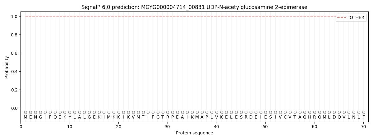
| Other | SP_Sec_SPI | LIPO_Sec_SPII | TAT_Tat_SPI | TATLIP_Sec_SPII | PILIN_Sec_SPIII |
|---|---|---|---|---|---|
| 1.000072 | 0.000000 | 0.000000 | 0.000000 | 0.000000 | 0.000000 |