You are browsing environment: HUMAN GUT
CAZyme Information: MGYG000004379_00602
You are here: Home > Sequence: MGYG000004379_00602
Basic Information |
Genomic context |
Full Sequence |
Enzyme annotations |
CAZy signature domains |
CDD domains |
CAZyme hits |
PDB hits |
Swiss-Prot hits |
SignalP and Lipop annotations |
TMHMM annotations
Basic Information help
| Species | CAG-628 sp000438415 | |||||||||||
|---|---|---|---|---|---|---|---|---|---|---|---|---|
| Lineage | Bacteria; Firmicutes; Bacilli; RF39; UBA660; CAG-628; CAG-628 sp000438415 | |||||||||||
| CAZyme ID | MGYG000004379_00602 | |||||||||||
| CAZy Family | GT2 | |||||||||||
| CAZyme Description | hypothetical protein | |||||||||||
| CAZyme Property |
|
|||||||||||
| Genome Property |
|
|||||||||||
| Gene Location | Start: 8285; End: 10264 Strand: + | |||||||||||
Full Sequence Download help
| MSKLNKIKTS IKEKGLIKTI KWLYESLIFR IKMIKRPSLD FAKIENTKDE NTYSYPKILP | 60 |
| NVFIVASIPY YDIGGGQRCS QLAKTFNKMG YNVIYLYGYK SSESKKFNLE MPVSSHMFIN | 120 |
| DKTILEVRNI VKNNDLFIFE APSLRFKDIL NIAISNKCKI VYENIDNWET SLGNDVFNES | 180 |
| MLISLLKNSD LLVGTAKPLV EQLNTYLEKY NIENKLAVYL PNAVDEELFC GLKELPKPKD | 240 |
| MITGEKTFLY YGSLWGSWFD WDLIKKLALH NENYSINIIG DYKNLTELIS TLPKNIHFLG | 300 |
| LKKQIELPNY LKYTDYSLIP FKTGEIGDFV SPLKIFEYIS MYTKVLSTTL PDIKGYPNLY | 360 |
| CGNTSDEWIN IVEKNYEVNK FDADNFIDNN SWHSRVTEII NNIYKEKNNS ILSGKLSIII | 420 |
| LNYNNKNIIF KSLNSLIKYN ELYNYELIVV DNGSTDGSYE ILKEKYSDKI KIIQNTKNGC | 480 |
| SSGRNLGVTL STKEYIMFLD SDQWVTNKYW LEPYENILKT DKNVGLIGWA AGFFNKEGYA | 540 |
| YHVVDSFPHK YMPANKLGRK DISYLGSGGM LLTRKLFDQI EGFDEKYDPT CYEDTDLSLK | 600 |
| VRNVNKEIVY CPYLGIIHLP HQTTDGGLLD HNSLFIEKRE YFKNKWTKQN ESLLKKHIK | 659 |
CAZyme Signature Domains help
| Family | Start | End | Evalue | family coverage |
|---|---|---|---|---|
| GT2 | 417 | 578 | 1.2e-24 | 0.9764705882352941 |
CDD Domains download full data without filtering help
| Cdd ID | Domain | E-Value | qStart | qEnd | sStart | sEnd | Domain Description |
|---|---|---|---|---|---|---|---|
| cd04186 | GT_2_like_c | 2.07e-34 | 418 | 619 | 1 | 165 | Subfamily of Glycosyltransferase Family GT2 of unknown function. GT-2 includes diverse families of glycosyltransferases with a common GT-A type structural fold, which has two tightly associated beta/alpha/beta domains that tend to form a continuous central sheet of at least eight beta-strands. These are enzymes that catalyze the transfer of sugar moieties from activated donor molecules to specific acceptor molecules, forming glycosidic bonds. Glycosyltransferases have been classified into more than 90 distinct sequence based families. |
| COG1216 | GT2 | 8.44e-25 | 415 | 648 | 4 | 250 | Glycosyltransferase, GT2 family [Carbohydrate transport and metabolism]. |
| cd00761 | Glyco_tranf_GTA_type | 6.11e-24 | 418 | 537 | 1 | 122 | Glycosyltransferase family A (GT-A) includes diverse families of glycosyl transferases with a common GT-A type structural fold. Glycosyltransferases (GTs) are enzymes that synthesize oligosaccharides, polysaccharides, and glycoconjugates by transferring the sugar moiety from an activated nucleotide-sugar donor to an acceptor molecule, which may be a growing oligosaccharide, a lipid, or a protein. Based on the stereochemistry of the donor and acceptor molecules, GTs are classified as either retaining or inverting enzymes. To date, all GT structures adopt one of two possible folds, termed GT-A fold and GT-B fold. This hierarchy includes diverse families of glycosyl transferases with a common GT-A type structural fold, which has two tightly associated beta/alpha/beta domains that tend to form a continuous central sheet of at least eight beta-strands. The majority of the proteins in this superfamily are Glycosyltransferase family 2 (GT-2) proteins. But it also includes families GT-43, GT-6, GT-8, GT13 and GT-7; which are evolutionarily related to GT-2 and share structure similarities. |
| pfam00535 | Glycos_transf_2 | 2.58e-21 | 417 | 575 | 1 | 161 | Glycosyl transferase family 2. Diverse family, transferring sugar from UDP-glucose, UDP-N-acetyl- galactosamine, GDP-mannose or CDP-abequose, to a range of substrates including cellulose, dolichol phosphate and teichoic acids. |
| cd04950 | GT4_TuaH-like | 3.62e-20 | 160 | 395 | 125 | 361 | teichuronic acid biosynthesis glycosyltransferase TuaH and similar proteins. Members of this family may function in teichuronic acid biosynthesis/cell wall biogenesis. Glycosyltransferases catalyze the transfer of sugar moieties from activated donor molecules to specific acceptor molecules, forming glycosidic bonds. The acceptor molecule can be a lipid, a protein, a heterocyclic compound, or another carbohydrate residue. This group of glycosyltransferases is most closely related to the previously defined glycosyltransferase family 1 (GT1). The members of this family may transfer UDP, ADP, GDP, or CMP linked sugars. The diverse enzymatic activities among members of this family reflect a wide range of biological functions. The protein structure available for this family has the GTB topology, one of the two protein topologies observed for nucleotide-sugar-dependent glycosyltransferases. GTB proteins have distinct N- and C- terminal domains each containing a typical Rossmann fold. The two domains have high structural homology despite minimal sequence homology. The large cleft that separates the two domains includes the catalytic center and permits a high degree of flexibility. |
CAZyme Hits help
| Hit ID | E-Value | Query Start | Query End | Hit Start | Hit End |
|---|---|---|---|---|---|
| ALS18183.1 | 1.58e-194 | 39 | 654 | 391 | 1001 |
| QCJ03883.1 | 1.58e-194 | 39 | 654 | 391 | 1001 |
| ANF15306.1 | 1.58e-194 | 39 | 654 | 391 | 1001 |
| AOR95255.1 | 1.58e-194 | 39 | 654 | 391 | 1001 |
| ALP91686.1 | 1.58e-194 | 39 | 654 | 391 | 1001 |
PDB Hits download full data without filtering help
| Hit ID | E-Value | Query Start | Query End | Hit Start | Hit End | Description |
|---|---|---|---|---|---|---|
| 5HEA_A | 5.19e-10 | 416 | 541 | 7 | 131 | CgTstructure in hexamer [Streptococcus parasanguinis FW213],5HEA_B CgT structure in hexamer [Streptococcus parasanguinis FW213],5HEA_C CgT structure in hexamer [Streptococcus parasanguinis FW213],5HEC_A CgT structure in dimer [Streptococcus parasanguinis FW213],5HEC_B CgT structure in dimer [Streptococcus parasanguinis FW213] |
| 2Z87_A | 4.56e-09 | 411 | 528 | 90 | 208 | Crystalstructure of chondroitin polymerase from Escherichia coli strain K4 (K4CP) complexed with UDP-GalNAc and UDP [Escherichia coli],2Z87_B Crystal structure of chondroitin polymerase from Escherichia coli strain K4 (K4CP) complexed with UDP-GalNAc and UDP [Escherichia coli] |
| 2Z86_A | 4.57e-09 | 411 | 528 | 91 | 209 | Crystalstructure of chondroitin polymerase from Escherichia coli strain K4 (K4CP) complexed with UDP-GlcUA and UDP [Escherichia coli],2Z86_B Crystal structure of chondroitin polymerase from Escherichia coli strain K4 (K4CP) complexed with UDP-GlcUA and UDP [Escherichia coli],2Z86_C Crystal structure of chondroitin polymerase from Escherichia coli strain K4 (K4CP) complexed with UDP-GlcUA and UDP [Escherichia coli],2Z86_D Crystal structure of chondroitin polymerase from Escherichia coli strain K4 (K4CP) complexed with UDP-GlcUA and UDP [Escherichia coli] |
| 6P61_A | 9.21e-08 | 416 | 630 | 15 | 214 | Structureof a Glycosyltransferase from Leptospira borgpetersenii serovar Hardjo-bovis (strain JB197) [Leptospira borgpetersenii serovar Hardjo-bovis str. JB197],6P61_B Structure of a Glycosyltransferase from Leptospira borgpetersenii serovar Hardjo-bovis (strain JB197) [Leptospira borgpetersenii serovar Hardjo-bovis str. JB197],6P61_C Structure of a Glycosyltransferase from Leptospira borgpetersenii serovar Hardjo-bovis (strain JB197) [Leptospira borgpetersenii serovar Hardjo-bovis str. JB197],6P61_D Structure of a Glycosyltransferase from Leptospira borgpetersenii serovar Hardjo-bovis (strain JB197) [Leptospira borgpetersenii serovar Hardjo-bovis str. JB197] |
| 5TZE_C | 3.09e-06 | 415 | 508 | 2 | 99 | Crystalstructure of S. aureus TarS in complex with UDP-GlcNAc [Staphylococcus aureus],5TZE_E Crystal structure of S. aureus TarS in complex with UDP-GlcNAc [Staphylococcus aureus],5TZI_C Crystal structure of S. aureus TarS 1-349 [Staphylococcus aureus],5TZJ_A Crystal structure of S. aureus TarS 1-349 in complex with UDP-GlcNAc [Staphylococcus aureus],5TZJ_C Crystal structure of S. aureus TarS 1-349 in complex with UDP-GlcNAc [Staphylococcus aureus],5TZK_C Crystal structure of S. aureus TarS 1-349 in complex with UDP [Staphylococcus aureus] |
Swiss-Prot Hits download full data without filtering help
| Hit ID | E-Value | Query Start | Query End | Hit Start | Hit End | Description |
|---|---|---|---|---|---|---|
| A0A0H2URH7 | 5.12e-12 | 411 | 610 | 2 | 207 | Glycosyltransferase GlyA OS=Streptococcus pneumoniae serotype 4 (strain ATCC BAA-334 / TIGR4) OX=170187 GN=glyA PE=3 SV=1 |
| Q8L0V4 | 2.64e-08 | 411 | 528 | 148 | 266 | Chondroitin synthase OS=Escherichia coli OX=562 GN=kfoC PE=1 SV=1 |
| P22639 | 1.11e-07 | 415 | 527 | 2 | 115 | Uncharacterized glycosyltransferase alr2836 OS=Nostoc sp. (strain PCC 7120 / SAG 25.82 / UTEX 2576) OX=103690 GN=alr2836 PE=3 SV=2 |
| P55465 | 1.17e-07 | 416 | 646 | 627 | 874 | Uncharacterized protein y4gI OS=Sinorhizobium fredii (strain NBRC 101917 / NGR234) OX=394 GN=NGR_a03550 PE=4 SV=1 |
| P71057 | 5.20e-07 | 416 | 509 | 6 | 100 | Putative glycosyltransferase EpsH OS=Bacillus subtilis (strain 168) OX=224308 GN=epsH PE=2 SV=1 |
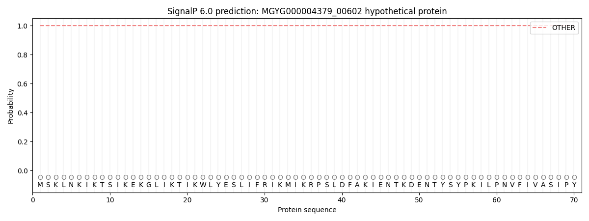
SignalP and Lipop Annotations help
This protein is predicted as OTHER
| Other | SP_Sec_SPI | LIPO_Sec_SPII | TAT_Tat_SPI | TATLIP_Sec_SPII | PILIN_Sec_SPIII |
|---|---|---|---|---|---|
| 0.999962 | 0.000055 | 0.000002 | 0.000000 | 0.000000 | 0.000001 |