You are browsing environment: HUMAN GUT
CAZyme Information: MGYG000001295_00218
You are here: Home > Sequence: MGYG000001295_00218
Basic Information |
Genomic context |
Full Sequence |
Enzyme annotations |
CAZy signature domains |
CDD domains |
CAZyme hits |
PDB hits |
Swiss-Prot hits |
SignalP and Lipop annotations |
TMHMM annotations
Basic Information help
| Species | Desulfovibrio fairfieldensis | |||||||||||
|---|---|---|---|---|---|---|---|---|---|---|---|---|
| Lineage | Bacteria; Desulfobacterota; Desulfovibrionia; Desulfovibrionales; Desulfovibrionaceae; Desulfovibrio; Desulfovibrio fairfieldensis | |||||||||||
| CAZyme ID | MGYG000001295_00218 | |||||||||||
| CAZy Family | GT2 | |||||||||||
| CAZyme Description | D-inositol-3-phosphate glycosyltransferase | |||||||||||
| CAZyme Property |
|
|||||||||||
| Genome Property |
|
|||||||||||
| Gene Location | Start: 21136; End: 23343 Strand: + | |||||||||||
Full Sequence Download help
| MLRISVIVPV YNTCSYLSKC LESLFSQTLC DIEIICVDDG STDGSSDVLR DFAQKDSRLK | 60 |
| VLKHERNKGA AAARNTGLAV ARGEYLGFVD SDDYVSNDYF EKLYIVAKEN TADIAKARYF | 120 |
| YEKKDDIQKE DKYNISIEEN KYNFSINFTT AIYRHSIIKN NKIRFPDGIT NYEDVVFLIM | 180 |
| CVCCANKIKT IDSVYYHYIK RTCSSTSKDY KKIFDMTLQA CFEIVFILNK TKIPYNNYNK | 240 |
| LFRQCINVLL SISVTPSDIS ENSAYKVIKL LENYKFLPQK IKKSKLPYTK RSVVEIFWDI | 300 |
| RRISAAPFFP KNDQKFIYTL YKMSHFISIC LFISTLAPGG AERQIVTLAK ALARKGVVVY | 360 |
| LVYYESKGNC GHYLGMLKNT YVIHFSYALN NTVVSGSSFC RKYQDLYHEI RAMGLPPLSI | 420 |
| LALTGALTFI SPDILHCYLD GNNILGGSVG ILGKVPGIVL SFRSVDPATA QEVYTDMALK | 480 |
| YYTFLLRYNN VALEANSNCG AYSYAHWLRI DLDRIVVNPN GIDSSLLNRE KFSSRQIARR | 540 |
| SLGIDESIPI VLFLGRYHAC KCPDVLLSVA DELRKKIPSV LFLVAGDGMQ HDAEIGFLLQ | 600 |
| QYKLTDNIRL LGPRKDVLNL LIAADVLLMT SKIEGFPNVV MEAMSAGRPV VATRVGAIPE | 660 |
| LVREGKDGFL HNVGDVVGLC ESLQFLLSDS KTRNRMGQNA KQRILEDFTS DRLAQRALLQ | 720 |
| YSSLLGQCQD EKEQV | 735 |
CAZyme Signature Domains help
| Family | Start | End | Evalue | family coverage |
|---|---|---|---|---|
| GT2 | 5 | 150 | 3.6e-38 | 0.8588235294117647 |
| GT4 | 544 | 697 | 2.1e-34 | 0.9625 |
CDD Domains download full data without filtering help
| Cdd ID | Domain | E-Value | qStart | qEnd | sStart | sEnd | Domain Description |
|---|---|---|---|---|---|---|---|
| cd03801 | GT4_PimA-like | 1.26e-55 | 331 | 716 | 3 | 360 | phosphatidyl-myo-inositol mannosyltransferase. This family is most closely related to the GT4 family of glycosyltransferases and named after PimA in Propionibacterium freudenreichii, which is involved in the biosynthesis of phosphatidyl-myo-inositol mannosides (PIM) which are early precursors in the biosynthesis of lipomannans (LM) and lipoarabinomannans (LAM), and catalyzes the addition of a mannosyl residue from GDP-D-mannose (GDP-Man) to the position 2 of the carrier lipid phosphatidyl-myo-inositol (PI) to generate a phosphatidyl-myo-inositol bearing an alpha-1,2-linked mannose residue (PIM1). Glycosyltransferases catalyze the transfer of sugar moieties from activated donor molecules to specific acceptor molecules, forming glycosidic bonds. The acceptor molecule can be a lipid, a protein, a heterocyclic compound, or another carbohydrate residue. This group of glycosyltransferases is most closely related to the previously defined glycosyltransferase family 1 (GT1). The members of this family may transfer UDP, ADP, GDP, or CMP linked sugars. The diverse enzymatic activities among members of this family reflect a wide range of biological functions. The protein structure available for this family has the GTB topology, one of the two protein topologies observed for nucleotide-sugar-dependent glycosyltransferases. GTB proteins have distinct N- and C- terminal domains each containing a typical Rossmann fold. The two domains have high structural homology despite minimal sequence homology. The large cleft that separates the two domains includes the catalytic center and permits a high degree of flexibility. The members of this family are found mainly in certain bacteria and archaea. |
| cd03807 | GT4_WbnK-like | 3.17e-54 | 333 | 725 | 6 | 361 | Shigella dysenteriae WbnK and similar proteins. This family is most closely related to the GT4 family of glycosyltransferases. WbnK in Shigella dysenteriae has been shown to be involved in the type 7 O-antigen biosynthesis. |
| cd03819 | GT4_WavL-like | 2.24e-45 | 331 | 700 | 2 | 332 | Vibrio cholerae WavL and similar sequences. This family is most closely related to the GT4 family of glycosyltransferases. WavL in Vibrio cholerae has been shown to be involved in the biosynthesis of the lipopolysaccharide core. |
| cd03808 | GT4_CapM-like | 1.11e-42 | 331 | 711 | 3 | 351 | capsular polysaccharide biosynthesis glycosyltransferase CapM and similar proteins. This family is most closely related to the GT4 family of glycosyltransferases. CapM in Staphylococcus aureus is required for the synthesis of type 1 capsular polysaccharides. |
| COG0438 | RfaB | 2.54e-40 | 327 | 727 | 3 | 380 | Glycosyltransferase involved in cell wall bisynthesis [Cell wall/membrane/envelope biogenesis]. |
CAZyme Hits help
| Hit ID | E-Value | Query Start | Query End | Hit Start | Hit End |
|---|---|---|---|---|---|
| QAZ69598.1 | 4.08e-67 | 301 | 726 | 63 | 493 |
| SLM50328.1 | 2.12e-47 | 333 | 725 | 71 | 463 |
| AMD90318.1 | 2.26e-39 | 4 | 195 | 4 | 193 |
| CCG55516.1 | 4.84e-39 | 1 | 213 | 2 | 228 |
| AGA66956.1 | 5.69e-38 | 1 | 213 | 1 | 227 |
PDB Hits download full data without filtering help
| Hit ID | E-Value | Query Start | Query End | Hit Start | Hit End | Description |
|---|---|---|---|---|---|---|
| 5HEA_A | 9.31e-26 | 4 | 151 | 7 | 148 | CgTstructure in hexamer [Streptococcus parasanguinis FW213],5HEA_B CgT structure in hexamer [Streptococcus parasanguinis FW213],5HEA_C CgT structure in hexamer [Streptococcus parasanguinis FW213],5HEC_A CgT structure in dimer [Streptococcus parasanguinis FW213],5HEC_B CgT structure in dimer [Streptococcus parasanguinis FW213] |
| 3BCV_A | 4.32e-25 | 3 | 198 | 6 | 224 | Crystalstructure of a putative glycosyltransferase from Bacteroides fragilis [Bacteroides fragilis NCTC 9343],3BCV_B Crystal structure of a putative glycosyltransferase from Bacteroides fragilis [Bacteroides fragilis NCTC 9343] |
| 3MBO_A | 1.35e-20 | 334 | 713 | 11 | 394 | CrystalStructure of the Glycosyltransferase BaBshA bound with UDP and L-malate [Bacillus anthracis],3MBO_B Crystal Structure of the Glycosyltransferase BaBshA bound with UDP and L-malate [Bacillus anthracis],3MBO_C Crystal Structure of the Glycosyltransferase BaBshA bound with UDP and L-malate [Bacillus anthracis],3MBO_D Crystal Structure of the Glycosyltransferase BaBshA bound with UDP and L-malate [Bacillus anthracis],3MBO_E Crystal Structure of the Glycosyltransferase BaBshA bound with UDP and L-malate [Bacillus anthracis],3MBO_F Crystal Structure of the Glycosyltransferase BaBshA bound with UDP and L-malate [Bacillus anthracis],3MBO_G Crystal Structure of the Glycosyltransferase BaBshA bound with UDP and L-malate [Bacillus anthracis],3MBO_H Crystal Structure of the Glycosyltransferase BaBshA bound with UDP and L-malate [Bacillus anthracis] |
| 2JJM_A | 1.47e-20 | 539 | 713 | 202 | 374 | CrystalStructure of a family GT4 glycosyltransferase from Bacillus anthracis ORF BA1558. [Bacillus anthracis str. Ames],2JJM_B Crystal Structure of a family GT4 glycosyltransferase from Bacillus anthracis ORF BA1558. [Bacillus anthracis str. Ames],2JJM_C Crystal Structure of a family GT4 glycosyltransferase from Bacillus anthracis ORF BA1558. [Bacillus anthracis str. Ames],2JJM_D Crystal Structure of a family GT4 glycosyltransferase from Bacillus anthracis ORF BA1558. [Bacillus anthracis str. Ames],2JJM_E Crystal Structure of a family GT4 glycosyltransferase from Bacillus anthracis ORF BA1558. [Bacillus anthracis str. Ames],2JJM_F Crystal Structure of a family GT4 glycosyltransferase from Bacillus anthracis ORF BA1558. [Bacillus anthracis str. Ames],2JJM_G Crystal Structure of a family GT4 glycosyltransferase from Bacillus anthracis ORF BA1558. [Bacillus anthracis str. Ames],2JJM_H Crystal Structure of a family GT4 glycosyltransferase from Bacillus anthracis ORF BA1558. [Bacillus anthracis str. Ames],2JJM_I Crystal Structure of a family GT4 glycosyltransferase from Bacillus anthracis ORF BA1558. [Bacillus anthracis str. Ames],2JJM_J Crystal Structure of a family GT4 glycosyltransferase from Bacillus anthracis ORF BA1558. [Bacillus anthracis str. Ames],2JJM_K Crystal Structure of a family GT4 glycosyltransferase from Bacillus anthracis ORF BA1558. [Bacillus anthracis str. Ames],2JJM_L Crystal Structure of a family GT4 glycosyltransferase from Bacillus anthracis ORF BA1558. [Bacillus anthracis str. Ames] |
| 3C4Q_A | 4.92e-19 | 339 | 700 | 22 | 382 | Structureof the retaining glycosyltransferase MshA : The first step in mycothiol biosynthesis. Organism : Corynebacterium glutamicum- Complex with UDP [Corynebacterium glutamicum],3C4Q_B Structure of the retaining glycosyltransferase MshA : The first step in mycothiol biosynthesis. Organism : Corynebacterium glutamicum- Complex with UDP [Corynebacterium glutamicum],3C4V_A Structure of the retaining glycosyltransferase MshA:The first step in mycothiol biosynthesis. Organism: Corynebacterium glutamicum : Complex with UDP and 1L-INS-1-P. [Corynebacterium glutamicum],3C4V_B Structure of the retaining glycosyltransferase MshA:The first step in mycothiol biosynthesis. Organism: Corynebacterium glutamicum : Complex with UDP and 1L-INS-1-P. [Corynebacterium glutamicum] |
Swiss-Prot Hits download full data without filtering help
| Hit ID | E-Value | Query Start | Query End | Hit Start | Hit End | Description |
|---|---|---|---|---|---|---|
| P71059 | 7.07e-30 | 4 | 290 | 5 | 307 | Uncharacterized glycosyltransferase EpsJ OS=Bacillus subtilis (strain 168) OX=224308 GN=epsJ PE=2 SV=1 |
| P71057 | 1.22e-27 | 4 | 218 | 6 | 231 | Putative glycosyltransferase EpsH OS=Bacillus subtilis (strain 168) OX=224308 GN=epsH PE=2 SV=1 |
| A0A0H2UR96 | 2.71e-26 | 4 | 246 | 5 | 270 | Glycosyltransferase GlyG OS=Streptococcus pneumoniae serotype 4 (strain ATCC BAA-334 / TIGR4) OX=170187 GN=glyG PE=1 SV=1 |
| E0U4V7 | 8.16e-26 | 2 | 201 | 1 | 207 | Poly(ribitol-phosphate) beta-glucosyltransferase OS=Bacillus spizizenii (strain ATCC 23059 / NRRL B-14472 / W23) OX=655816 GN=tarQ PE=1 SV=1 |
| A0A0H2URH7 | 2.38e-25 | 3 | 270 | 6 | 286 | Glycosyltransferase GlyA OS=Streptococcus pneumoniae serotype 4 (strain ATCC BAA-334 / TIGR4) OX=170187 GN=glyA PE=3 SV=1 |
SignalP and Lipop Annotations help
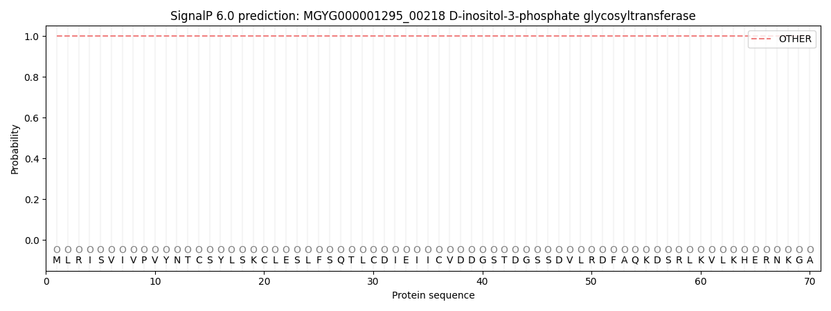
This protein is predicted as OTHER
| Other | SP_Sec_SPI | LIPO_Sec_SPII | TAT_Tat_SPI | TATLIP_Sec_SPII | PILIN_Sec_SPIII |
|---|---|---|---|---|---|
| 1.000085 | 0.000000 | 0.000000 | 0.000000 | 0.000000 | 0.000000 |