You are browsing environment: HUMAN GUT
CAZyme Information: MGYG000002846_00456
You are here: Home > Sequence: MGYG000002846_00456
Basic Information |
Genomic context |
Full Sequence |
Enzyme annotations |
CAZy signature domains |
CDD domains |
CAZyme hits |
PDB hits |
Swiss-Prot hits |
SignalP and Lipop annotations |
TMHMM annotations
Basic Information help
| Species | Desulfovibrio sp900540515 | |||||||||||
|---|---|---|---|---|---|---|---|---|---|---|---|---|
| Lineage | Bacteria; Desulfobacterota; Desulfovibrionia; Desulfovibrionales; Desulfovibrionaceae; Desulfovibrio; Desulfovibrio sp900540515 | |||||||||||
| CAZyme ID | MGYG000002846_00456 | |||||||||||
| CAZy Family | GT51 | |||||||||||
| CAZyme Description | Penicillin-binding protein 1C | |||||||||||
| CAZyme Property |
|
|||||||||||
| Genome Property |
|
|||||||||||
| Gene Location | Start: 34110; End: 36413 Strand: - | |||||||||||
Full Sequence Download help
| MGLPLLLCCA ALAFALALYA TPRPDIYGRA GFSRSFLDRQ GRVLRITLAP DGAYRIFTPL | 60 |
| TALPPELIEA TLLYEDRFFF RHPGVNPLSL LRSAVSMVLG GRRMGGSTIS MQVARLSGKF | 120 |
| STASIPGKLR QIWHALALER HYDKDEILEA YLNLAPYGAN VEGAGAAARV WFHKEAGELS | 180 |
| LPEILALAPV PQHPAARNPL TSRGRALSQA RARLDKIWRQ AHPRAAGQGL PDVPLRVHGP | 240 |
| SELPFAAPHA VDSLLAAPDL PQGDIVTTLD LGLQRLLERQ LRRAVEAGRL WGMDNAAALL | 300 |
| LDWSSGEILA LAGSADYFNA AIQGQVDGTA ARRSPGSTLK PFIYALALQE GLIHPESLLS | 360 |
| DTPRVFKGYE PENADGGFRG PVSARMALLG SRNIPAISLA GQLPAPGLYG FLRKAGVRFE | 420 |
| HGPEHYGLSL VLGGAEVSMR ELAGLYAVLP NRGVWRAPML RRDEAAPEPL PLLRAEAAFV | 480 |
| ALQMLRAPSP QGFDRTPVYW KTGTSNGLRD AWTAGIFGPY VLVVWVGRFD GASNPGFNGL | 540 |
| HAAAPVFFSI QRALEAQSPP GAPADPGANA DGLKVRRIAV CAATGDTDLS LCPEPSRQTG | 600 |
| TWFIPGVSPI RDSGILRRIL VDEVTGFRQC RAVPGRTREV VWEFWPADLR RLFTQAGVRK | 660 |
| PAALPLAPEC RDTDAPWQVP GRAPLISSPK SGLIYTASLG NPQKIPLLAD ADADADCVYW | 720 |
| FAGARYLGRS APDEPLFWQA APGVTRLTAV DDLGRSSSVR VVTESVP | 767 |
CAZyme Signature Domains help
| Family | Start | End | Evalue | family coverage |
|---|---|---|---|---|
| GT51 | 54 | 215 | 2.1e-42 | 0.9152542372881356 |
CDD Domains download full data without filtering help
| Cdd ID | Domain | E-Value | qStart | qEnd | sStart | sEnd | Domain Description |
|---|---|---|---|---|---|---|---|
| TIGR02073 | PBP_1c | 0.0 | 33 | 764 | 11 | 727 | penicillin-binding protein 1C. This subfamily of the penicillin binding proteins includes the member from E. coli designated penicillin-binding protein 1C. Members have both transglycosylase and transpeptidase domains and are involved in forming cross-links in the late stages of peptidoglycan biosynthesis. All members of this subfamily are presumed to have the same basic function. [Cell envelope, Biosynthesis and degradation of murein sacculus and peptidoglycan] |
| COG4953 | PbpC | 0.0 | 32 | 761 | 35 | 729 | Membrane carboxypeptidase/penicillin-binding protein PbpC [Cell wall/membrane/envelope biogenesis]. |
| TIGR02074 | PBP_1a_fam | 1.31e-101 | 54 | 547 | 1 | 521 | penicillin-binding protein, 1A family. Bacterial that synthesize a cell wall of peptidoglycan (murein) generally have several transglycosylases and transpeptidases for the task. This family consists of bifunctional transglycosylase/transpeptidase penicillin-binding proteins (PBP). In the Proteobacteria, this family includes PBP 1A but not the paralogous PBP 1B (TIGR02071). This family also includes related proteins, often designated PBP 1A, from other bacterial lineages. [Cell envelope, Biosynthesis and degradation of murein sacculus and peptidoglycan] |
| COG0744 | MrcB | 1.00e-100 | 36 | 601 | 58 | 646 | Membrane carboxypeptidase (penicillin-binding protein) [Cell wall/membrane/envelope biogenesis]. |
| PRK11240 | PRK11240 | 3.45e-98 | 51 | 681 | 58 | 698 | penicillin-binding protein 1C; Provisional |
CAZyme Hits help
| Hit ID | E-Value | Query Start | Query End | Hit Start | Hit End |
|---|---|---|---|---|---|
| AMD89899.1 | 0.0 | 2 | 767 | 15 | 780 |
| QTO41888.1 | 1.14e-250 | 13 | 767 | 28 | 771 |
| QCC84882.1 | 4.17e-249 | 10 | 761 | 51 | 791 |
| SPD36822.1 | 2.33e-237 | 31 | 767 | 46 | 772 |
| ATD81196.1 | 2.33e-237 | 31 | 767 | 46 | 772 |
PDB Hits download full data without filtering help
| Hit ID | E-Value | Query Start | Query End | Hit Start | Hit End | Description |
|---|---|---|---|---|---|---|
| 4OON_A | 7.48e-29 | 63 | 530 | 47 | 696 | Crystalstructure of PBP1a in complex with compound 17 ((4Z,8S,11E,14S)-5-(2-amino-1,3-thiazol-4-yl)-14-(5,6-dihydroxy-1,3-dioxo-1,3-dihydro-2H-isoindol-2-yl)-8-formyl-2-methyl-6-oxo-3,10-dioxa-4,7,11-triazatetradeca-4,11-diene-2,12,14-tricarboxylic acid) [Pseudomonas aeruginosa PAO1] |
| 3UDF_A | 4.54e-28 | 63 | 550 | 47 | 711 | ChainA, Penicillin-binding protein 1a [Acinetobacter baumannii],3UDF_B Chain B, Penicillin-binding protein 1a [Acinetobacter baumannii],3UDI_A Chain A, Penicillin-binding protein 1a [Acinetobacter baumannii],3UDI_B Chain B, Penicillin-binding protein 1a [Acinetobacter baumannii],3UDX_A Chain A, Penicillin-binding protein 1a [Acinetobacter baumannii],3UDX_B Chain B, Penicillin-binding protein 1a [Acinetobacter baumannii],3UE0_A Chain A, Penicillin-binding protein 1a [Acinetobacter baumannii],3UE0_B Chain B, Penicillin-binding protein 1a [Acinetobacter baumannii],3UE1_A Chain A, Penicillin-binding protein 1a [Acinetobacter baumannii],3UE1_B Chain B, Penicillin-binding protein 1a [Acinetobacter baumannii] |
| 2JE5_A | 1.06e-23 | 66 | 559 | 65 | 638 | StructuralAnd Mechanistic Basis Of Penicillin Binding Protein Inhibition By Lactivicins [Streptococcus pneumoniae R6],2JE5_B Structural And Mechanistic Basis Of Penicillin Binding Protein Inhibition By Lactivicins [Streptococcus pneumoniae R6] |
| 3NB6_A | 2.03e-22 | 19 | 222 | 4 | 188 | Crystalstructure of Aquifex aeolicus peptidoglycan glycosyltransferase in complex with Methylphosphoryl Neryl Moenomycin [Aquifex aeolicus] |
| 2OQO_A | 2.76e-22 | 19 | 222 | 4 | 188 | Crystalstructure of a peptidoglycan glycosyltransferase from a class A PBP: insight into bacterial cell wall synthesis [Aquifex aeolicus VF5],3D3H_A Crystal structure of a complex of the peptidoglycan glycosyltransferase domain from Aquifex aeolicus and neryl moenomycin A [Aquifex aeolicus],3NB7_A Crystal structure of Aquifex Aeolicus Peptidoglycan Glycosyltransferase in complex with Decarboxylated Neryl Moenomycin [Aquifex aeolicus] |
Swiss-Prot Hits download full data without filtering help
| Hit ID | E-Value | Query Start | Query End | Hit Start | Hit End | Description |
|---|---|---|---|---|---|---|
| P76577 | 1.69e-83 | 51 | 762 | 60 | 768 | Penicillin-binding protein 1C OS=Escherichia coli (strain K12) OX=83333 GN=pbpC PE=1 SV=1 |
| Q9KUC0 | 3.88e-39 | 55 | 530 | 180 | 691 | Penicillin-binding protein 1B OS=Vibrio cholerae serotype O1 (strain ATCC 39315 / El Tor Inaba N16961) OX=243277 GN=mrcB PE=3 SV=1 |
| Q9PGD4 | 1.80e-34 | 63 | 552 | 78 | 752 | Penicillin-binding protein 1A OS=Xylella fastidiosa (strain 9a5c) OX=160492 GN=mrcA PE=3 SV=2 |
| O66874 | 1.28e-33 | 55 | 559 | 63 | 672 | Penicillin-binding protein 1A OS=Aquifex aeolicus (strain VF5) OX=224324 GN=mrcA PE=1 SV=1 |
| P38050 | 2.15e-33 | 60 | 527 | 72 | 569 | Penicillin-binding protein 1F OS=Bacillus subtilis (strain 168) OX=224308 GN=pbpF PE=2 SV=2 |
SignalP and Lipop Annotations help
This protein is predicted as OTHER
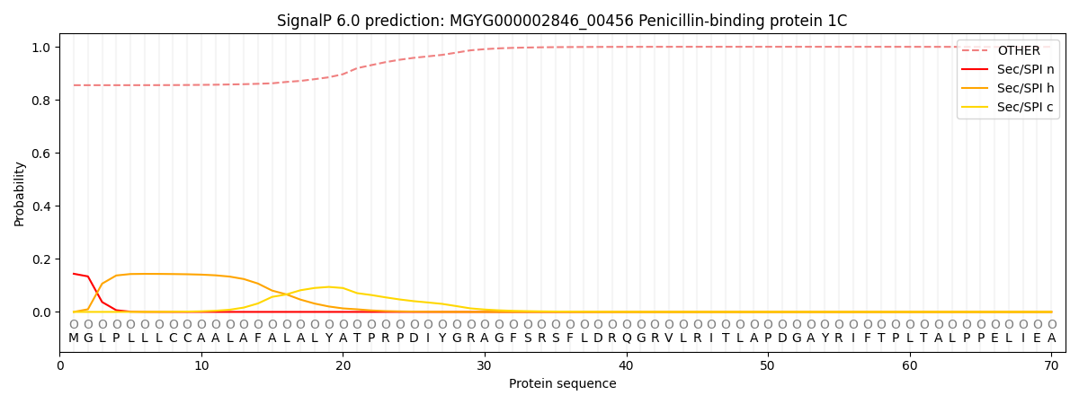
| Other | SP_Sec_SPI | LIPO_Sec_SPII | TAT_Tat_SPI | TATLIP_Sec_SPII | PILIN_Sec_SPIII |
|---|---|---|---|---|---|
| 0.861432 | 0.136786 | 0.001305 | 0.000148 | 0.000112 | 0.000224 |