You are browsing environment: HUMAN GUT
CAZyme Information: MGYG000000012_01433
You are here: Home > Sequence: MGYG000000012_01433
Basic Information |
Genomic context |
Full Sequence |
Enzyme annotations |
CAZy signature domains |
CDD domains |
CAZyme hits |
PDB hits |
Swiss-Prot hits |
SignalP and Lipop annotations |
TMHMM annotations
Basic Information help
| Species | Bacillus subtilis | |||||||||||
|---|---|---|---|---|---|---|---|---|---|---|---|---|
| Lineage | Bacteria; Firmicutes; Bacilli; Bacillales; Bacillaceae; Bacillus; Bacillus subtilis | |||||||||||
| CAZyme ID | MGYG000000012_01433 | |||||||||||
| CAZy Family | GT4 | |||||||||||
| CAZyme Description | Putative glycosyltransferase EpsD | |||||||||||
| CAZyme Property |
|
|||||||||||
| Genome Property |
|
|||||||||||
| Gene Location | Start: 356286; End: 357431 Strand: - | |||||||||||
CDD Domains download full data without filtering help
| Cdd ID | Domain | E-Value | qStart | qEnd | sStart | sEnd | Domain Description |
|---|---|---|---|---|---|---|---|
| cd03808 | GT4_CapM-like | 2.33e-113 | 4 | 364 | 1 | 357 | capsular polysaccharide biosynthesis glycosyltransferase CapM and similar proteins. This family is most closely related to the GT4 family of glycosyltransferases. CapM in Staphylococcus aureus is required for the synthesis of type 1 capsular polysaccharides. |
| cd03801 | GT4_PimA-like | 1.38e-62 | 4 | 368 | 1 | 365 | phosphatidyl-myo-inositol mannosyltransferase. This family is most closely related to the GT4 family of glycosyltransferases and named after PimA in Propionibacterium freudenreichii, which is involved in the biosynthesis of phosphatidyl-myo-inositol mannosides (PIM) which are early precursors in the biosynthesis of lipomannans (LM) and lipoarabinomannans (LAM), and catalyzes the addition of a mannosyl residue from GDP-D-mannose (GDP-Man) to the position 2 of the carrier lipid phosphatidyl-myo-inositol (PI) to generate a phosphatidyl-myo-inositol bearing an alpha-1,2-linked mannose residue (PIM1). Glycosyltransferases catalyze the transfer of sugar moieties from activated donor molecules to specific acceptor molecules, forming glycosidic bonds. The acceptor molecule can be a lipid, a protein, a heterocyclic compound, or another carbohydrate residue. This group of glycosyltransferases is most closely related to the previously defined glycosyltransferase family 1 (GT1). The members of this family may transfer UDP, ADP, GDP, or CMP linked sugars. The diverse enzymatic activities among members of this family reflect a wide range of biological functions. The protein structure available for this family has the GTB topology, one of the two protein topologies observed for nucleotide-sugar-dependent glycosyltransferases. GTB proteins have distinct N- and C- terminal domains each containing a typical Rossmann fold. The two domains have high structural homology despite minimal sequence homology. The large cleft that separates the two domains includes the catalytic center and permits a high degree of flexibility. The members of this family are found mainly in certain bacteria and archaea. |
| pfam00534 | Glycos_transf_1 | 6.97e-53 | 196 | 350 | 1 | 157 | Glycosyl transferases group 1. Mutations in this domain of PIGA lead to disease (Paroxysmal Nocturnal haemoglobinuria). Members of this family transfer activated sugars to a variety of substrates, including glycogen, Fructose-6-phosphate and lipopolysaccharides. Members of this family transfer UDP, ADP, GDP or CMP linked sugars. The eukaryotic glycogen synthases may be distant members of this family. |
| COG0438 | RfaB | 2.07e-47 | 27 | 372 | 31 | 378 | Glycosyltransferase involved in cell wall bisynthesis [Cell wall/membrane/envelope biogenesis]. |
| cd03819 | GT4_WavL-like | 1.44e-40 | 27 | 364 | 25 | 341 | Vibrio cholerae WavL and similar sequences. This family is most closely related to the GT4 family of glycosyltransferases. WavL in Vibrio cholerae has been shown to be involved in the biosynthesis of the lipopolysaccharide core. |
CAZyme Hits help
| Hit ID | E-Value | Query Start | Query End | Hit Start | Hit End |
|---|---|---|---|---|---|
| QRZ92300.1 | 2.83e-284 | 1 | 381 | 1 | 381 |
| ASB62516.1 | 8.11e-284 | 1 | 381 | 1 | 381 |
| AFI30011.1 | 4.69e-283 | 1 | 381 | 1 | 381 |
| AUS11547.1 | 1.34e-282 | 1 | 381 | 1 | 381 |
| AUZ40503.1 | 7.46e-280 | 1 | 381 | 1 | 381 |
PDB Hits download full data without filtering help
| Hit ID | E-Value | Query Start | Query End | Hit Start | Hit End | Description |
|---|---|---|---|---|---|---|
| 6N1X_A | 1.72e-17 | 59 | 323 | 69 | 325 | ChainA, Glycosyltransferase [Staphylococcus aureus subsp. aureus CN1] |
| 6D9T_A | 2.02e-17 | 59 | 323 | 85 | 341 | BshAfrom Staphylococcus aureus complexed with UDP [Staphylococcus aureus] |
| 5D00_A | 6.79e-15 | 59 | 369 | 70 | 373 | Crystalstructure of BshA from B. subtilis complexed with N-acetylglucosaminyl-malate and UMP [Bacillus subtilis subsp. subtilis str. 168],5D00_B Crystal structure of BshA from B. subtilis complexed with N-acetylglucosaminyl-malate and UMP [Bacillus subtilis subsp. subtilis str. 168],5D01_A Crystal structure of BshA from B. subtilis complexed with N-acetylglucosaminyl-malate [Bacillus subtilis subsp. subtilis str. 168],5D01_B Crystal structure of BshA from B. subtilis complexed with N-acetylglucosaminyl-malate [Bacillus subtilis subsp. subtilis str. 168] |
| 7EC2_A | 1.05e-14 | 204 | 356 | 325 | 478 | ChainA, Glycosyl transferase, group 1 family protein [Staphylococcus aureus subsp. aureus USA300],7EC2_B Chain B, Glycosyl transferase, group 1 family protein [Staphylococcus aureus subsp. aureus USA300] |
| 3C4Q_A | 1.57e-13 | 170 | 369 | 196 | 403 | Structureof the retaining glycosyltransferase MshA : The first step in mycothiol biosynthesis. Organism : Corynebacterium glutamicum- Complex with UDP [Corynebacterium glutamicum],3C4Q_B Structure of the retaining glycosyltransferase MshA : The first step in mycothiol biosynthesis. Organism : Corynebacterium glutamicum- Complex with UDP [Corynebacterium glutamicum],3C4V_A Structure of the retaining glycosyltransferase MshA:The first step in mycothiol biosynthesis. Organism: Corynebacterium glutamicum : Complex with UDP and 1L-INS-1-P. [Corynebacterium glutamicum],3C4V_B Structure of the retaining glycosyltransferase MshA:The first step in mycothiol biosynthesis. Organism: Corynebacterium glutamicum : Complex with UDP and 1L-INS-1-P. [Corynebacterium glutamicum] |
Swiss-Prot Hits download full data without filtering help
| Hit ID | E-Value | Query Start | Query End | Hit Start | Hit End | Description |
|---|---|---|---|---|---|---|
| P71053 | 4.03e-269 | 1 | 381 | 1 | 381 | Putative glycosyltransferase EpsD OS=Bacillus subtilis (strain 168) OX=224308 GN=epsD PE=2 SV=1 |
| Q9R9N1 | 5.90e-18 | 215 | 351 | 186 | 318 | Lipopolysaccharide core biosynthesis glycosyltransferase LpsE OS=Rhizobium meliloti (strain 1021) OX=266834 GN=lpsE PE=3 SV=1 |
| C9ZH13 | 1.49e-15 | 79 | 372 | 129 | 433 | D-inositol 3-phosphate glycosyltransferase OS=Streptomyces scabiei (strain 87.22) OX=680198 GN=mshA PE=3 SV=1 |
| Q65CC7 | 4.88e-15 | 82 | 368 | 88 | 381 | Alpha-D-kanosaminyltransferase OS=Streptomyces kanamyceticus OX=1967 GN=kanE PE=1 SV=1 |
| Q9FCG5 | 6.70e-15 | 79 | 370 | 129 | 443 | D-inositol 3-phosphate glycosyltransferase OS=Streptomyces coelicolor (strain ATCC BAA-471 / A3(2) / M145) OX=100226 GN=mshA PE=2 SV=2 |
SignalP and Lipop Annotations help
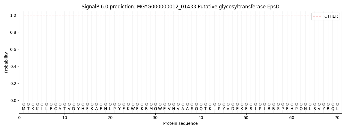
This protein is predicted as OTHER
| Other | SP_Sec_SPI | LIPO_Sec_SPII | TAT_Tat_SPI | TATLIP_Sec_SPII | PILIN_Sec_SPIII |
|---|---|---|---|---|---|
| 1.000069 | 0.000000 | 0.000000 | 0.000000 | 0.000000 | 0.000000 |