You are browsing environment: HUMAN GUT
CAZyme Information: MGYG000000066_03173
You are here: Home > Sequence: MGYG000000066_03173
Basic Information |
Genomic context |
Full Sequence |
Enzyme annotations |
CAZy signature domains |
CDD domains |
CAZyme hits |
PDB hits |
Swiss-Prot hits |
SignalP and Lipop annotations |
TMHMM annotations
Basic Information help
| Species | GCA-900066755 sp900066755 | |||||||||||
|---|---|---|---|---|---|---|---|---|---|---|---|---|
| Lineage | Bacteria; Firmicutes_A; Clostridia; Lachnospirales; Lachnospiraceae; GCA-900066755; GCA-900066755 sp900066755 | |||||||||||
| CAZyme ID | MGYG000000066_03173 | |||||||||||
| CAZy Family | GT2 | |||||||||||
| CAZyme Description | hypothetical protein | |||||||||||
| CAZyme Property |
|
|||||||||||
| Genome Property |
|
|||||||||||
| Gene Location | Start: 9270; End: 10742 Strand: - | |||||||||||
CAZyme Signature Domains help
| Family | Start | End | Evalue | family coverage |
|---|---|---|---|---|
| GT2 | 66 | 173 | 8.8e-29 | 0.6588235294117647 |
CDD Domains download full data without filtering help
| Cdd ID | Domain | E-Value | qStart | qEnd | sStart | sEnd | Domain Description |
|---|---|---|---|---|---|---|---|
| cd04184 | GT2_RfbC_Mx_like | 1.51e-105 | 65 | 261 | 3 | 201 | Myxococcus xanthus RfbC like proteins are required for O-antigen biosynthesis. The rfbC gene encodes a predicted protein of 1,276 amino acids, which is required for O-antigen biosynthesis in Myxococcus xanthus. It is a subfamily of Glycosyltransferase Family GT2, which includes diverse families of glycosyl transferases with a common GT-A type structural fold, which has two tightly associated beta/alpha/beta domains that tend to form a continuous central sheet of at least eight beta-strands. These are enzymes that catalyze the transfer of sugar moieties from activated donor molecules to specific acceptor molecules, forming glycosidic bonds. |
| cd06433 | GT_2_WfgS_like | 1.28e-36 | 66 | 267 | 1 | 200 | WfgS and WfeV are involved in O-antigen biosynthesis. Escherichia coli WfgS and Shigella dysenteriae WfeV are glycosyltransferase 2 family enzymes involved in O-antigen biosynthesis. GT-2 enzymes have GT-A type structural fold, which has two tightly associated beta/alpha/beta domains that tend to form a continuous central sheet of at least eight beta-strands. These are enzymes that catalyze the transfer of sugar moieties from activated donor molecules to specific acceptor molecules, forming glycosidic bonds. Glycosyltransferases have been classified into more than 90 distinct sequence based families. |
| pfam00535 | Glycos_transf_2 | 5.61e-29 | 66 | 172 | 1 | 108 | Glycosyl transferase family 2. Diverse family, transferring sugar from UDP-glucose, UDP-N-acetyl- galactosamine, GDP-mannose or CDP-abequose, to a range of substrates including cellulose, dolichol phosphate and teichoic acids. |
| cd00761 | Glyco_tranf_GTA_type | 2.15e-23 | 67 | 252 | 1 | 156 | Glycosyltransferase family A (GT-A) includes diverse families of glycosyl transferases with a common GT-A type structural fold. Glycosyltransferases (GTs) are enzymes that synthesize oligosaccharides, polysaccharides, and glycoconjugates by transferring the sugar moiety from an activated nucleotide-sugar donor to an acceptor molecule, which may be a growing oligosaccharide, a lipid, or a protein. Based on the stereochemistry of the donor and acceptor molecules, GTs are classified as either retaining or inverting enzymes. To date, all GT structures adopt one of two possible folds, termed GT-A fold and GT-B fold. This hierarchy includes diverse families of glycosyl transferases with a common GT-A type structural fold, which has two tightly associated beta/alpha/beta domains that tend to form a continuous central sheet of at least eight beta-strands. The majority of the proteins in this superfamily are Glycosyltransferase family 2 (GT-2) proteins. But it also includes families GT-43, GT-6, GT-8, GT13 and GT-7; which are evolutionarily related to GT-2 and share structure similarities. |
| cd04196 | GT_2_like_d | 2.37e-20 | 66 | 263 | 1 | 205 | Subfamily of Glycosyltransferase Family GT2 of unknown function. GT-2 includes diverse families of glycosyltransferases with a common GT-A type structural fold, which has two tightly associated beta/alpha/beta domains that tend to form a continuous central sheet of at least eight beta-strands. These are enzymes that catalyze the transfer of sugar moieties from activated donor molecules to specific acceptor molecules, forming glycosidic bonds. Glycosyltransferases have been classified into more than 90 distinct sequence based families. |
CAZyme Hits help
| Hit ID | E-Value | Query Start | Query End | Hit Start | Hit End |
|---|---|---|---|---|---|
| QNM10257.1 | 6.50e-117 | 5 | 486 | 18 | 550 |
| QCU01342.1 | 1.40e-116 | 5 | 472 | 24 | 550 |
| QIB58022.1 | 3.21e-115 | 5 | 472 | 20 | 546 |
| QMW77454.1 | 3.21e-115 | 5 | 472 | 20 | 546 |
| QBE99393.1 | 6.39e-115 | 5 | 472 | 20 | 546 |
PDB Hits download full data without filtering help
| Hit ID | E-Value | Query Start | Query End | Hit Start | Hit End | Description |
|---|---|---|---|---|---|---|
| 2Z86_A | 3.88e-15 | 66 | 268 | 378 | 579 | Crystalstructure of chondroitin polymerase from Escherichia coli strain K4 (K4CP) complexed with UDP-GlcUA and UDP [Escherichia coli],2Z86_B Crystal structure of chondroitin polymerase from Escherichia coli strain K4 (K4CP) complexed with UDP-GlcUA and UDP [Escherichia coli],2Z86_C Crystal structure of chondroitin polymerase from Escherichia coli strain K4 (K4CP) complexed with UDP-GlcUA and UDP [Escherichia coli],2Z86_D Crystal structure of chondroitin polymerase from Escherichia coli strain K4 (K4CP) complexed with UDP-GlcUA and UDP [Escherichia coli] |
| 2Z87_A | 3.88e-15 | 66 | 268 | 377 | 578 | Crystalstructure of chondroitin polymerase from Escherichia coli strain K4 (K4CP) complexed with UDP-GalNAc and UDP [Escherichia coli],2Z87_B Crystal structure of chondroitin polymerase from Escherichia coli strain K4 (K4CP) complexed with UDP-GalNAc and UDP [Escherichia coli] |
| 5HEA_A | 6.30e-08 | 66 | 155 | 8 | 97 | CgTstructure in hexamer [Streptococcus parasanguinis FW213],5HEA_B CgT structure in hexamer [Streptococcus parasanguinis FW213],5HEA_C CgT structure in hexamer [Streptococcus parasanguinis FW213],5HEC_A CgT structure in dimer [Streptococcus parasanguinis FW213],5HEC_B CgT structure in dimer [Streptococcus parasanguinis FW213] |
Swiss-Prot Hits download full data without filtering help
| Hit ID | E-Value | Query Start | Query End | Hit Start | Hit End | Description |
|---|---|---|---|---|---|---|
| P55465 | 7.48e-57 | 58 | 473 | 361 | 841 | Uncharacterized protein y4gI OS=Sinorhizobium fredii (strain NBRC 101917 / NGR234) OX=394 GN=NGR_a03550 PE=4 SV=1 |
| Q50864 | 7.97e-36 | 30 | 486 | 286 | 794 | O-antigen biosynthesis protein RfbC OS=Myxococcus xanthus OX=34 GN=rfbC PE=4 SV=1 |
| Q9CMP0 | 2.42e-17 | 66 | 268 | 436 | 637 | Chondroitin synthase OS=Pasteurella multocida (strain Pm70) OX=272843 GN=fcbD PE=3 SV=1 |
| Q7BLV3 | 2.43e-17 | 66 | 268 | 443 | 644 | Hyaluronan synthase OS=Pasteurella multocida OX=747 GN=hyaD PE=1 SV=2 |
| Q8L0V4 | 2.29e-14 | 66 | 268 | 435 | 636 | Chondroitin synthase OS=Escherichia coli OX=562 GN=kfoC PE=1 SV=1 |
SignalP and Lipop Annotations help
This protein is predicted as OTHER
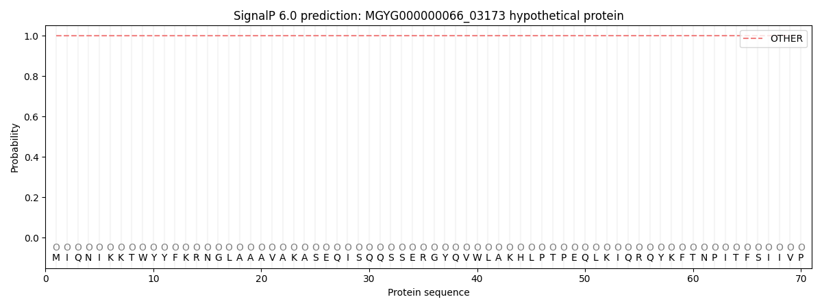
| Other | SP_Sec_SPI | LIPO_Sec_SPII | TAT_Tat_SPI | TATLIP_Sec_SPII | PILIN_Sec_SPIII |
|---|---|---|---|---|---|
| 0.999825 | 0.000174 | 0.000018 | 0.000001 | 0.000001 | 0.000006 |