You are browsing environment: HUMAN GUT
CAZyme Information: MGYG000000110_02138
You are here: Home > Sequence: MGYG000000110_02138
Basic Information |
Genomic context |
Full Sequence |
Enzyme annotations |
CAZy signature domains |
CDD domains |
CAZyme hits |
PDB hits |
Swiss-Prot hits |
SignalP and Lipop annotations |
TMHMM annotations
Basic Information help
| Species | Kluyvera sp902363335 | |||||||||||
|---|---|---|---|---|---|---|---|---|---|---|---|---|
| Lineage | Bacteria; Proteobacteria; Gammaproteobacteria; Enterobacterales; Enterobacteriaceae; Kluyvera; Kluyvera sp902363335 | |||||||||||
| CAZyme ID | MGYG000000110_02138 | |||||||||||
| CAZy Family | GT4 | |||||||||||
| CAZyme Description | Lipopolysaccharide core biosynthesis protein RfaG | |||||||||||
| CAZyme Property |
|
|||||||||||
| Genome Property |
|
|||||||||||
| Gene Location | Start: 211795; End: 212919 Strand: + | |||||||||||
CDD Domains download full data without filtering help
| Cdd ID | Domain | E-Value | qStart | qEnd | sStart | sEnd | Domain Description |
|---|---|---|---|---|---|---|---|
| cd03801 | GT4_PimA-like | 3.20e-46 | 3 | 368 | 2 | 362 | phosphatidyl-myo-inositol mannosyltransferase. This family is most closely related to the GT4 family of glycosyltransferases and named after PimA in Propionibacterium freudenreichii, which is involved in the biosynthesis of phosphatidyl-myo-inositol mannosides (PIM) which are early precursors in the biosynthesis of lipomannans (LM) and lipoarabinomannans (LAM), and catalyzes the addition of a mannosyl residue from GDP-D-mannose (GDP-Man) to the position 2 of the carrier lipid phosphatidyl-myo-inositol (PI) to generate a phosphatidyl-myo-inositol bearing an alpha-1,2-linked mannose residue (PIM1). Glycosyltransferases catalyze the transfer of sugar moieties from activated donor molecules to specific acceptor molecules, forming glycosidic bonds. The acceptor molecule can be a lipid, a protein, a heterocyclic compound, or another carbohydrate residue. This group of glycosyltransferases is most closely related to the previously defined glycosyltransferase family 1 (GT1). The members of this family may transfer UDP, ADP, GDP, or CMP linked sugars. The diverse enzymatic activities among members of this family reflect a wide range of biological functions. The protein structure available for this family has the GTB topology, one of the two protein topologies observed for nucleotide-sugar-dependent glycosyltransferases. GTB proteins have distinct N- and C- terminal domains each containing a typical Rossmann fold. The two domains have high structural homology despite minimal sequence homology. The large cleft that separates the two domains includes the catalytic center and permits a high degree of flexibility. The members of this family are found mainly in certain bacteria and archaea. |
| pfam00534 | Glycos_transf_1 | 1.03e-37 | 196 | 350 | 2 | 157 | Glycosyl transferases group 1. Mutations in this domain of PIGA lead to disease (Paroxysmal Nocturnal haemoglobinuria). Members of this family transfer activated sugars to a variety of substrates, including glycogen, Fructose-6-phosphate and lipopolysaccharides. Members of this family transfer UDP, ADP, GDP or CMP linked sugars. The eukaryotic glycogen synthases may be distant members of this family. |
| COG0438 | RfaB | 1.59e-29 | 1 | 352 | 1 | 357 | Glycosyltransferase involved in cell wall bisynthesis [Cell wall/membrane/envelope biogenesis]. |
| cd03800 | GT4_sucrose_synthase | 5.44e-24 | 147 | 365 | 173 | 394 | sucrose-phosphate synthase and similar proteins. This family is most closely related to the GT4 family of glycosyltransferases. The sucrose-phosphate synthases in this family may be unique to plants and photosynthetic bacteria. This enzyme catalyzes the synthesis of sucrose 6-phosphate from fructose 6-phosphate and uridine 5'-diphosphate-glucose, a key regulatory step of sucrose metabolism. The activity of this enzyme is regulated by phosphorylation and moderated by the concentration of various metabolites and light. |
| cd03798 | GT4_WlbH-like | 4.66e-22 | 160 | 353 | 173 | 358 | Bordetella parapertussis WlbH and similar proteins. This family is most closely related to the GT4 family of glycosyltransferases. Staphylococcus aureus CapJ may be involved in capsule polysaccharide biosynthesis. WlbH in Bordetella parapertussis has been shown to be required for the biosynthesis of a trisaccharide that, when attached to the B. pertussis lipopolysaccharide (LPS) core (band B), generates band A LPS. |
CAZyme Hits help
| Hit ID | E-Value | Query Start | Query End | Hit Start | Hit End |
|---|---|---|---|---|---|
| QGH37270.1 | 1.59e-266 | 1 | 374 | 1 | 374 |
| QGH28288.1 | 1.59e-266 | 1 | 374 | 1 | 374 |
| VDZ85725.1 | 9.20e-266 | 1 | 374 | 1 | 374 |
| BBR18701.1 | 9.24e-250 | 1 | 374 | 1 | 374 |
| BBS89581.1 | 9.24e-250 | 1 | 374 | 1 | 374 |
PDB Hits download full data without filtering help
| Hit ID | E-Value | Query Start | Query End | Hit Start | Hit End | Description |
|---|---|---|---|---|---|---|
| 2IW1_A | 5.55e-234 | 1 | 374 | 1 | 374 | CrystalStructure of WaaG, a glycosyltransferase involved in lipopolysaccharide biosynthesis [Escherichia coli str. K-12 substr. W3110] |
| 2IV7_A | 4.16e-229 | 2 | 374 | 2 | 374 | CrystalStructure of WaaG, a glycosyltransferase involved in lipopolysaccharide biosynthesis [Escherichia coli str. K-12 substr. W3110] |
| 3C4Q_A | 6.47e-11 | 140 | 367 | 168 | 398 | Structureof the retaining glycosyltransferase MshA : The first step in mycothiol biosynthesis. Organism : Corynebacterium glutamicum- Complex with UDP [Corynebacterium glutamicum],3C4Q_B Structure of the retaining glycosyltransferase MshA : The first step in mycothiol biosynthesis. Organism : Corynebacterium glutamicum- Complex with UDP [Corynebacterium glutamicum],3C4V_A Structure of the retaining glycosyltransferase MshA:The first step in mycothiol biosynthesis. Organism: Corynebacterium glutamicum : Complex with UDP and 1L-INS-1-P. [Corynebacterium glutamicum],3C4V_B Structure of the retaining glycosyltransferase MshA:The first step in mycothiol biosynthesis. Organism: Corynebacterium glutamicum : Complex with UDP and 1L-INS-1-P. [Corynebacterium glutamicum] |
| 3C48_A | 6.67e-11 | 140 | 367 | 188 | 418 | Structureof the retaining glycosyltransferase MshA: The first step in mycothiol biosynthesis. Organism: Corynebacterium glutamicum- APO (OPEN) structure. [Corynebacterium glutamicum],3C48_B Structure of the retaining glycosyltransferase MshA: The first step in mycothiol biosynthesis. Organism: Corynebacterium glutamicum- APO (OPEN) structure. [Corynebacterium glutamicum] |
| 2N58_A | 9.68e-11 | 103 | 132 | 1 | 30 | Structureof an N-terminal membrane-anchoring region of the glycosyltransferase WaaG [Escherichia coli K-12] |
Swiss-Prot Hits download full data without filtering help
| Hit ID | E-Value | Query Start | Query End | Hit Start | Hit End | Description |
|---|---|---|---|---|---|---|
| P25740 | 3.04e-233 | 1 | 374 | 1 | 374 | Lipopolysaccharide core biosynthesis protein RfaG OS=Escherichia coli (strain K12) OX=83333 GN=rfaG PE=1 SV=1 |
| Q82G92 | 1.18e-13 | 139 | 367 | 212 | 447 | D-inositol 3-phosphate glycosyltransferase OS=Streptomyces avermitilis (strain ATCC 31267 / DSM 46492 / JCM 5070 / NBRC 14893 / NCIMB 12804 / NRRL 8165 / MA-4680) OX=227882 GN=mshA PE=3 SV=1 |
| D5USX8 | 2.02e-12 | 139 | 352 | 180 | 401 | D-inositol 3-phosphate glycosyltransferase OS=Tsukamurella paurometabola (strain ATCC 8368 / DSM 20162 / CCUG 35730 / CIP 100753 / JCM 10117 / KCTC 9821 / NBRC 16120 / NCIMB 702349 / NCTC 13040) OX=521096 GN=mshA PE=3 SV=1 |
| Q65CC7 | 4.12e-11 | 136 | 348 | 149 | 360 | Alpha-D-kanosaminyltransferase OS=Streptomyces kanamyceticus OX=1967 GN=kanE PE=1 SV=1 |
| C9ZH13 | 6.61e-11 | 139 | 319 | 193 | 381 | D-inositol 3-phosphate glycosyltransferase OS=Streptomyces scabiei (strain 87.22) OX=680198 GN=mshA PE=3 SV=1 |
SignalP and Lipop Annotations help
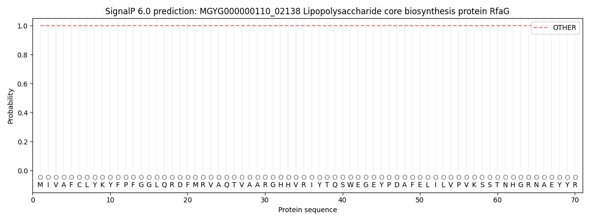
This protein is predicted as OTHER
| Other | SP_Sec_SPI | LIPO_Sec_SPII | TAT_Tat_SPI | TATLIP_Sec_SPII | PILIN_Sec_SPIII |
|---|---|---|---|---|---|
| 1.000044 | 0.000000 | 0.000000 | 0.000000 | 0.000000 | 0.000000 |