You are browsing environment: HUMAN GUT
CAZyme Information: MGYG000000117_01431
You are here: Home > Sequence: MGYG000000117_01431
Basic Information |
Genomic context |
Full Sequence |
Enzyme annotations |
CAZy signature domains |
CDD domains |
CAZyme hits |
PDB hits |
Swiss-Prot hits |
SignalP and Lipop annotations |
TMHMM annotations
Basic Information help
| Species | Barnesiella sp003150885 | |||||||||||
|---|---|---|---|---|---|---|---|---|---|---|---|---|
| Lineage | Bacteria; Bacteroidota; Bacteroidia; Bacteroidales; Barnesiellaceae; Barnesiella; Barnesiella sp003150885 | |||||||||||
| CAZyme ID | MGYG000000117_01431 | |||||||||||
| CAZy Family | GT2 | |||||||||||
| CAZyme Description | Dodecaprenyl-phosphate galacturonate synthase | |||||||||||
| CAZyme Property |
|
|||||||||||
| Genome Property |
|
|||||||||||
| Gene Location | Start: 157391; End: 158104 Strand: - | |||||||||||
CAZyme Signature Domains help
| Family | Start | End | Evalue | family coverage |
|---|---|---|---|---|
| GT2 | 7 | 164 | 1.6e-33 | 0.9176470588235294 |
CDD Domains download full data without filtering help
| Cdd ID | Domain | E-Value | qStart | qEnd | sStart | sEnd | Domain Description |
|---|---|---|---|---|---|---|---|
| cd04179 | DPM_DPG-synthase_like | 1.19e-47 | 8 | 192 | 1 | 184 | DPM_DPG-synthase_like is a member of the Glycosyltransferase 2 superfamily. DPM1 is the catalytic subunit of eukaryotic dolichol-phosphate mannose (DPM) synthase. DPM synthase is required for synthesis of the glycosylphosphatidylinositol (GPI) anchor, N-glycan precursor, protein O-mannose, and C-mannose. In higher eukaryotes,the enzyme has three subunits, DPM1, DPM2 and DPM3. DPM is synthesized from dolichol phosphate and GDP-Man on the cytosolic surface of the ER membrane by DPM synthase and then is flipped onto the luminal side and used as a donor substrate. In lower eukaryotes, such as Saccharomyces cerevisiae and Trypanosoma brucei, DPM synthase consists of a single component (Dpm1p and TbDpm1, respectively) that possesses one predicted transmembrane region near the C terminus for anchoring to the ER membrane. In contrast, the Dpm1 homologues of higher eukaryotes, namely fission yeast, fungi, and animals, have no transmembrane region, suggesting the existence of adapter molecules for membrane anchoring. This family also includes bacteria and archaea DPM1_like enzymes. However, the enzyme structure and mechanism of function are not well understood. The UDP-glucose:dolichyl-phosphate glucosyltransferase (DPG_synthase) is a transmembrane-bound enzyme of the endoplasmic reticulum involved in protein N-linked glycosylation. This enzyme catalyzes the transfer of glucose from UDP-glucose to dolichyl phosphate. This protein family belongs to Glycosyltransferase 2 superfamily. |
| cd06442 | DPM1_like | 1.84e-32 | 8 | 232 | 1 | 223 | DPM1_like represents putative enzymes similar to eukaryotic DPM1. Proteins similar to eukaryotic DPM1, including enzymes from bacteria and archaea; DPM1 is the catalytic subunit of eukaryotic dolichol-phosphate mannose (DPM) synthase. DPM synthase is required for synthesis of the glycosylphosphatidylinositol (GPI) anchor, N-glycan precursor, protein O-mannose, and C-mannose. In higher eukaryotes,the enzyme has three subunits, DPM1, DPM2 and DPM3. DPM is synthesized from dolichol phosphate and GDP-Man on the cytosolic surface of the ER membrane by DPM synthase and then is flipped onto the luminal side and used as a donor substrate. In lower eukaryotes, such as Saccharomyces cerevisiae and Trypanosoma brucei, DPM synthase consists of a single component (Dpm1p and TbDpm1, respectively) that possesses one predicted transmembrane region near the C terminus for anchoring to the ER membrane. In contrast, the Dpm1 homologues of higher eukaryotes, namely fission yeast, fungi, and animals, have no transmembrane region, suggesting the existence of adapter molecules for membrane anchoring. This family also includes bacteria and archaea DPM1_like enzymes. However, the enzyme structure and mechanism of function are not well understood. This protein family belongs to Glycosyltransferase 2 superfamily. |
| pfam00535 | Glycos_transf_2 | 3.21e-31 | 7 | 168 | 1 | 159 | Glycosyl transferase family 2. Diverse family, transferring sugar from UDP-glucose, UDP-N-acetyl- galactosamine, GDP-mannose or CDP-abequose, to a range of substrates including cellulose, dolichol phosphate and teichoic acids. |
| cd04188 | DPG_synthase | 6.65e-31 | 8 | 205 | 1 | 202 | DPG_synthase is involved in protein N-linked glycosylation. UDP-glucose:dolichyl-phosphate glucosyltransferase (DPG_synthase) is a transmembrane-bound enzyme of the endoplasmic reticulum involved in protein N-linked glycosylation. This enzyme catalyzes the transfer of glucose from UDP-glucose to dolichyl phosphate. |
| cd00761 | Glyco_tranf_GTA_type | 3.96e-22 | 8 | 129 | 1 | 118 | Glycosyltransferase family A (GT-A) includes diverse families of glycosyl transferases with a common GT-A type structural fold. Glycosyltransferases (GTs) are enzymes that synthesize oligosaccharides, polysaccharides, and glycoconjugates by transferring the sugar moiety from an activated nucleotide-sugar donor to an acceptor molecule, which may be a growing oligosaccharide, a lipid, or a protein. Based on the stereochemistry of the donor and acceptor molecules, GTs are classified as either retaining or inverting enzymes. To date, all GT structures adopt one of two possible folds, termed GT-A fold and GT-B fold. This hierarchy includes diverse families of glycosyl transferases with a common GT-A type structural fold, which has two tightly associated beta/alpha/beta domains that tend to form a continuous central sheet of at least eight beta-strands. The majority of the proteins in this superfamily are Glycosyltransferase family 2 (GT-2) proteins. But it also includes families GT-43, GT-6, GT-8, GT13 and GT-7; which are evolutionarily related to GT-2 and share structure similarities. |
CAZyme Hits help
| Hit ID | E-Value | Query Start | Query End | Hit Start | Hit End |
|---|---|---|---|---|---|
| SNV40053.1 | 1.39e-106 | 6 | 237 | 4 | 235 |
| ADY33467.1 | 1.39e-106 | 6 | 237 | 4 | 235 |
| AZS32053.1 | 1.77e-102 | 1 | 235 | 1 | 235 |
| AHF12392.1 | 1.61e-95 | 4 | 235 | 6 | 237 |
| AFK05056.1 | 2.44e-93 | 6 | 237 | 3 | 226 |
PDB Hits download full data without filtering help
| Hit ID | E-Value | Query Start | Query End | Hit Start | Hit End | Description |
|---|---|---|---|---|---|---|
| 5MLZ_A | 2.68e-14 | 6 | 233 | 25 | 249 | Dolichylphosphate mannose synthase in complex with GDP and Mg2+ [Pyrococcus furiosus DSM 3638],5MM0_A Dolichyl phosphate mannose synthase in complex with GDP-mannose and Mn2+ (donor complex) [Pyrococcus furiosus DSM 3638],5MM1_A Dolichyl phosphate mannose synthase in complex with GDP and dolichyl phosphate mannose [Pyrococcus furiosus DSM 3638] |
| 6YV7_B | 2.32e-10 | 6 | 106 | 44 | 143 | MannosyltransferasePcManGT from Pyrobaculum calidifontis [Pyrobaculum calidifontis JCM 11548],6YV8_B Mannosyltransferase PcManGT from Pyrobaculum calidifontis in complex with GDP and Mn2+ [Pyrobaculum calidifontis JCM 11548],6YV9_A Mannosyltransferase PcManGT from Pyrobaculum calidifontis in complex with GDP-Man and Mn2+ [Pyrobaculum calidifontis JCM 11548] |
| 6YV7_A | 2.32e-10 | 6 | 106 | 45 | 144 | MannosyltransferasePcManGT from Pyrobaculum calidifontis [Pyrobaculum calidifontis JCM 11548],6YV8_A Mannosyltransferase PcManGT from Pyrobaculum calidifontis in complex with GDP and Mn2+ [Pyrobaculum calidifontis JCM 11548],6YV9_B Mannosyltransferase PcManGT from Pyrobaculum calidifontis in complex with GDP-Man and Mn2+ [Pyrobaculum calidifontis JCM 11548] |
| 4Y6N_A | 7.89e-09 | 5 | 100 | 48 | 141 | Crystalstructure of glucosyl-3-phosphoglycerate synthase from Mycobacterium tuberculosis in complex with Mn2+, uridine-diphosphate-glucose (UDP-Glc) and phosphoglyceric acid (PGA) - GpgS Mn2+ UDP-Glc PGA-1 [Mycobacterium tuberculosis H37Rv],4Y6U_A Mycobacterial protein [Mycobacterium tuberculosis H37Rv],4Y7F_A Crystal structure of glucosyl-3-phosphoglycerate synthase from Mycobacterium tuberculosis in complex with Mn2+, uridine-diphosphate-glucose (UDP-Glc) and 3-(phosphonooxy)propanoic acid (PPA) - GpgS Mn2+ UDP-Glc PPA [Mycobacterium tuberculosis H37Rv],4Y7G_A Crystal structure of glucosyl-3-phosphoglycerate synthase from Mycobacterium tuberculosis in complex with Mn2+, uridine-diphosphate-glucose (UDP-Glc) and glycerol 3-phosphate (G3P) - GpgS Mn2+ UDP-Glc G3P [Mycobacterium tuberculosis H37Rv],4Y9X_A Crystal structure of glucosyl-3-phosphoglycerate synthase from Mycobacterium tuberculosis in complex with Mn2+, uridine-diphosphate-glucose (UDP-Glc) and phosphoglyceric acid (PGA) - GpgS Mn2+ UDP-Glc PGA-3 [Mycobacterium tuberculosis H37Rv],5JQX_A Crystal structure of glucosyl-3-phosphoglycerate synthase from Mycobacterium tuberculosis in complex with phosphoglyceric acid (PGA) - GpgS*PGA [Mycobacterium tuberculosis H37Ra],5JQX_B Crystal structure of glucosyl-3-phosphoglycerate synthase from Mycobacterium tuberculosis in complex with phosphoglyceric acid (PGA) - GpgS*PGA [Mycobacterium tuberculosis H37Ra],5JQX_C Crystal structure of glucosyl-3-phosphoglycerate synthase from Mycobacterium tuberculosis in complex with phosphoglyceric acid (PGA) - GpgS*PGA [Mycobacterium tuberculosis H37Ra],5JQX_D Crystal structure of glucosyl-3-phosphoglycerate synthase from Mycobacterium tuberculosis in complex with phosphoglyceric acid (PGA) - GpgS*PGA [Mycobacterium tuberculosis H37Ra],5JSX_A Crystal structure of glucosyl-3-phosphoglycerate synthase from Mycobacterium tuberculosis in complex with Mn2+ and uridine-diphosphate-glucose (UDP-Glc) [Mycobacterium tuberculosis H37Ra],5JT0_A Crystal structure of glucosyl-3-phosphoglycerate synthase from Mycobacterium tuberculosis in complex with Mn2+, uridine-diphosphate (UDP) and glucosyl-3-phosphoglycerate (GPG) - GpgS*GPG*UDP*Mn2+ [Mycobacterium tuberculosis H37Rv],5JUC_A Crystal structure of glucosyl-3-phosphoglycerate synthase from Mycobacterium tuberculosis in complex with Mn2+, uridine-diphosphate (UDP) and glucosyl-3-phosphoglycerate (GPG) - GpgS*GPG*UDP*Mn2+_2 [Mycobacterium tuberculosis H37Rv],5JUD_A Crystal structure of glucosyl-3-phosphoglycerate synthase from Mycobacterium tuberculosis in complex with uridine-diphosphate (UDP) - GpgS*UDP [Mycobacterium tuberculosis variant bovis AF2122/97] |
| 3E25_A | 8.10e-09 | 5 | 100 | 44 | 137 | ChainA, Crystal structure of M. tuberculosis glucosyl-3-phosphoglycerate synthase [Mycobacterium tuberculosis],3E26_A Chain A, Crystal structure of M. tuberculosis glucosyl-3-phosphoglycerate synthase [Mycobacterium tuberculosis] |
Swiss-Prot Hits download full data without filtering help
| Hit ID | E-Value | Query Start | Query End | Hit Start | Hit End | Description |
|---|---|---|---|---|---|---|
| Q9LM93 | 2.05e-20 | 4 | 216 | 13 | 226 | Dolichol-phosphate mannosyltransferase subunit 1 OS=Arabidopsis thaliana OX=3702 GN=DPMS1 PE=1 SV=1 |
| Q54J42 | 9.20e-19 | 6 | 235 | 76 | 316 | Dolichyl-phosphate beta-glucosyltransferase OS=Dictyostelium discoideum OX=44689 GN=alg5 PE=2 SV=1 |
| A5GFZ5 | 1.80e-16 | 7 | 216 | 27 | 239 | Dolichol-phosphate mannosyltransferase subunit 1 OS=Sus scrofa OX=9823 GN=DPM1 PE=3 SV=1 |
| Q58619 | 5.20e-16 | 6 | 231 | 19 | 238 | Uncharacterized protein MJ1222 OS=Methanocaldococcus jannaschii (strain ATCC 43067 / DSM 2661 / JAL-1 / JCM 10045 / NBRC 100440) OX=243232 GN=MJ1222 PE=4 SV=1 |
| A2EK20 | 1.48e-15 | 2 | 232 | 71 | 309 | Dolichyl-phosphate beta-glucosyltransferase ALG5B OS=Trichomonas vaginalis OX=5722 GN=ALG5B PE=1 SV=1 |
SignalP and Lipop Annotations help
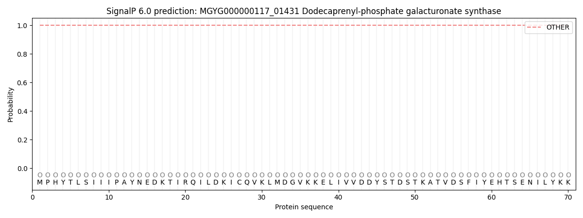
This protein is predicted as OTHER
| Other | SP_Sec_SPI | LIPO_Sec_SPII | TAT_Tat_SPI | TATLIP_Sec_SPII | PILIN_Sec_SPIII |
|---|---|---|---|---|---|
| 1.000061 | 0.000000 | 0.000000 | 0.000000 | 0.000000 | 0.000000 |