You are browsing environment: HUMAN GUT
CAZyme Information: MGYG000000132_02380
You are here: Home > Sequence: MGYG000000132_02380
Basic Information |
Genomic context |
Full Sequence |
Enzyme annotations |
CAZy signature domains |
CDD domains |
CAZyme hits |
PDB hits |
Swiss-Prot hits |
SignalP and Lipop annotations |
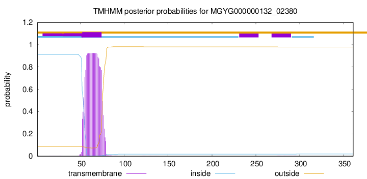
TMHMM annotations
Basic Information help
| Species | Beduini sp902363625 | |||||||||||
|---|---|---|---|---|---|---|---|---|---|---|---|---|
| Lineage | Bacteria; Firmicutes; Bacilli; Erysipelotrichales; Erysipelatoclostridiaceae; Beduini; Beduini sp902363625 | |||||||||||
| CAZyme ID | MGYG000000132_02380 | |||||||||||
| CAZy Family | GT4 | |||||||||||
| CAZyme Description | D-inositol-3-phosphate glycosyltransferase | |||||||||||
| CAZyme Property |
|
|||||||||||
| Genome Property |
|
|||||||||||
| Gene Location | Start: 119258; End: 120343 Strand: - | |||||||||||
CDD Domains download full data without filtering help
| Cdd ID | Domain | E-Value | qStart | qEnd | sStart | sEnd | Domain Description |
|---|---|---|---|---|---|---|---|
| cd03801 | GT4_PimA-like | 2.09e-42 | 3 | 361 | 1 | 366 | phosphatidyl-myo-inositol mannosyltransferase. This family is most closely related to the GT4 family of glycosyltransferases and named after PimA in Propionibacterium freudenreichii, which is involved in the biosynthesis of phosphatidyl-myo-inositol mannosides (PIM) which are early precursors in the biosynthesis of lipomannans (LM) and lipoarabinomannans (LAM), and catalyzes the addition of a mannosyl residue from GDP-D-mannose (GDP-Man) to the position 2 of the carrier lipid phosphatidyl-myo-inositol (PI) to generate a phosphatidyl-myo-inositol bearing an alpha-1,2-linked mannose residue (PIM1). Glycosyltransferases catalyze the transfer of sugar moieties from activated donor molecules to specific acceptor molecules, forming glycosidic bonds. The acceptor molecule can be a lipid, a protein, a heterocyclic compound, or another carbohydrate residue. This group of glycosyltransferases is most closely related to the previously defined glycosyltransferase family 1 (GT1). The members of this family may transfer UDP, ADP, GDP, or CMP linked sugars. The diverse enzymatic activities among members of this family reflect a wide range of biological functions. The protein structure available for this family has the GTB topology, one of the two protein topologies observed for nucleotide-sugar-dependent glycosyltransferases. GTB proteins have distinct N- and C- terminal domains each containing a typical Rossmann fold. The two domains have high structural homology despite minimal sequence homology. The large cleft that separates the two domains includes the catalytic center and permits a high degree of flexibility. The members of this family are found mainly in certain bacteria and archaea. |
| COG0438 | RfaB | 1.78e-34 | 3 | 361 | 1 | 375 | Glycosyltransferase involved in cell wall bisynthesis [Cell wall/membrane/envelope biogenesis]. |
| cd03798 | GT4_WlbH-like | 5.83e-34 | 4 | 315 | 1 | 329 | Bordetella parapertussis WlbH and similar proteins. This family is most closely related to the GT4 family of glycosyltransferases. Staphylococcus aureus CapJ may be involved in capsule polysaccharide biosynthesis. WlbH in Bordetella parapertussis has been shown to be required for the biosynthesis of a trisaccharide that, when attached to the B. pertussis lipopolysaccharide (LPS) core (band B), generates band A LPS. |
| cd03808 | GT4_CapM-like | 2.08e-30 | 22 | 344 | 15 | 344 | capsular polysaccharide biosynthesis glycosyltransferase CapM and similar proteins. This family is most closely related to the GT4 family of glycosyltransferases. CapM in Staphylococcus aureus is required for the synthesis of type 1 capsular polysaccharides. |
| pfam00534 | Glycos_transf_1 | 1.68e-29 | 186 | 335 | 4 | 149 | Glycosyl transferases group 1. Mutations in this domain of PIGA lead to disease (Paroxysmal Nocturnal haemoglobinuria). Members of this family transfer activated sugars to a variety of substrates, including glycogen, Fructose-6-phosphate and lipopolysaccharides. Members of this family transfer UDP, ADP, GDP or CMP linked sugars. The eukaryotic glycogen synthases may be distant members of this family. |
CAZyme Hits help
| Hit ID | E-Value | Query Start | Query End | Hit Start | Hit End |
|---|---|---|---|---|---|
| ALU13847.1 | 1.85e-108 | 1 | 361 | 4 | 365 |
| BCL58939.1 | 4.46e-108 | 1 | 360 | 1 | 357 |
| QQV05330.1 | 9.09e-106 | 1 | 360 | 1 | 359 |
| AYQ25818.1 | 2.83e-85 | 1 | 361 | 1 | 358 |
| QCQ14526.1 | 2.83e-85 | 1 | 361 | 1 | 358 |
PDB Hits download full data without filtering help
| Hit ID | E-Value | Query Start | Query End | Hit Start | Hit End | Description |
|---|---|---|---|---|---|---|
| 4X6L_A | 6.72e-11 | 187 | 338 | 321 | 472 | ChainA, TarM [Staphylococcus aureus subsp. aureus 21178],4X6L_B Chain B, TarM [Staphylococcus aureus subsp. aureus 21178],4X6L_C Chain C, TarM [Staphylococcus aureus subsp. aureus 21178],4X6L_D Chain D, TarM [Staphylococcus aureus subsp. aureus 21178],4X7P_A Chain A, TarM [Staphylococcus aureus subsp. aureus 21178],4X7P_B Chain B, TarM [Staphylococcus aureus subsp. aureus 21178] |
| 4X7M_A | 6.72e-11 | 187 | 338 | 321 | 472 | ChainA, TarM [Staphylococcus aureus subsp. aureus 21178],4X7M_B Chain B, TarM [Staphylococcus aureus subsp. aureus 21178],4X7R_A Chain A, TarM [Staphylococcus aureus subsp. aureus 21178],4X7R_B Chain B, TarM [Staphylococcus aureus subsp. aureus 21178] |
| 4WAC_A | 6.77e-11 | 187 | 338 | 326 | 477 | CrystalStructure of TarM [Staphylococcus aureus],4WAD_A Crystal Structure of TarM with UDP-GlcNAc [Staphylococcus aureus] |
| 3C4Q_A | 2.45e-08 | 128 | 360 | 165 | 402 | Structureof the retaining glycosyltransferase MshA : The first step in mycothiol biosynthesis. Organism : Corynebacterium glutamicum- Complex with UDP [Corynebacterium glutamicum],3C4Q_B Structure of the retaining glycosyltransferase MshA : The first step in mycothiol biosynthesis. Organism : Corynebacterium glutamicum- Complex with UDP [Corynebacterium glutamicum],3C4V_A Structure of the retaining glycosyltransferase MshA:The first step in mycothiol biosynthesis. Organism: Corynebacterium glutamicum : Complex with UDP and 1L-INS-1-P. [Corynebacterium glutamicum],3C4V_B Structure of the retaining glycosyltransferase MshA:The first step in mycothiol biosynthesis. Organism: Corynebacterium glutamicum : Complex with UDP and 1L-INS-1-P. [Corynebacterium glutamicum] |
| 3C48_A | 2.51e-08 | 128 | 360 | 185 | 422 | Structureof the retaining glycosyltransferase MshA: The first step in mycothiol biosynthesis. Organism: Corynebacterium glutamicum- APO (OPEN) structure. [Corynebacterium glutamicum],3C48_B Structure of the retaining glycosyltransferase MshA: The first step in mycothiol biosynthesis. Organism: Corynebacterium glutamicum- APO (OPEN) structure. [Corynebacterium glutamicum] |
Swiss-Prot Hits download full data without filtering help
| Hit ID | E-Value | Query Start | Query End | Hit Start | Hit End | Description |
|---|---|---|---|---|---|---|
| D5USX8 | 6.90e-15 | 128 | 343 | 178 | 394 | D-inositol 3-phosphate glycosyltransferase OS=Tsukamurella paurometabola (strain ATCC 8368 / DSM 20162 / CCUG 35730 / CIP 100753 / JCM 10117 / KCTC 9821 / NBRC 16120 / NCIMB 702349 / NCTC 13040) OX=521096 GN=mshA PE=3 SV=1 |
| A1SP12 | 9.88e-15 | 69 | 361 | 140 | 448 | D-inositol 3-phosphate glycosyltransferase OS=Nocardioides sp. (strain ATCC BAA-499 / JS614) OX=196162 GN=mshA PE=3 SV=1 |
| B8HCF8 | 6.80e-14 | 69 | 317 | 106 | 360 | D-inositol 3-phosphate glycosyltransferase OS=Pseudarthrobacter chlorophenolicus (strain ATCC 700700 / DSM 12829 / CIP 107037 / JCM 12360 / KCTC 9906 / NCIMB 13794 / A6) OX=452863 GN=mshA PE=3 SV=1 |
| A1R8N8 | 8.70e-14 | 64 | 361 | 95 | 397 | D-inositol 3-phosphate glycosyltransferase OS=Paenarthrobacter aurescens (strain TC1) OX=290340 GN=mshA PE=3 SV=1 |
| A3PU84 | 1.31e-13 | 124 | 347 | 174 | 392 | D-inositol 3-phosphate glycosyltransferase OS=Mycobacterium sp. (strain JLS) OX=164757 GN=mshA PE=3 SV=1 |
SignalP and Lipop Annotations help
This protein is predicted as OTHER

| Other | SP_Sec_SPI | LIPO_Sec_SPII | TAT_Tat_SPI | TATLIP_Sec_SPII | PILIN_Sec_SPIII |
|---|---|---|---|---|---|
| 0.999964 | 0.000027 | 0.000000 | 0.000000 | 0.000000 | 0.000000 |