You are browsing environment: HUMAN GUT
CAZyme Information: MGYG000000145_03496
You are here: Home > Sequence: MGYG000000145_03496
Basic Information |
Genomic context |
Full Sequence |
Enzyme annotations |
CAZy signature domains |
CDD domains |
CAZyme hits |
PDB hits |
Swiss-Prot hits |
SignalP and Lipop annotations |
TMHMM annotations
Basic Information help
| Species | Lacrimispora sp902363735 | |||||||||||
|---|---|---|---|---|---|---|---|---|---|---|---|---|
| Lineage | Bacteria; Firmicutes_A; Clostridia; Lachnospirales; Lachnospiraceae; Lacrimispora; Lacrimispora sp902363735 | |||||||||||
| CAZyme ID | MGYG000000145_03496 | |||||||||||
| CAZy Family | CBM50 | |||||||||||
| CAZyme Description | hypothetical protein | |||||||||||
| CAZyme Property |
|
|||||||||||
| Genome Property |
|
|||||||||||
| Gene Location | Start: 187081; End: 188553 Strand: + | |||||||||||
CDD Domains download full data without filtering help
| Cdd ID | Domain | E-Value | qStart | qEnd | sStart | sEnd | Domain Description |
|---|---|---|---|---|---|---|---|
| smart00257 | LysM | 3.47e-14 | 166 | 208 | 2 | 44 | Lysin motif. |
| pfam01476 | LysM | 2.58e-13 | 166 | 209 | 1 | 43 | LysM domain. The LysM (lysin motif) domain is about 40 residues long. It is found in a variety of enzymes involved in bacterial cell wall degradation. This domain may have a general peptidoglycan binding function. The structure of this domain is known. |
| smart00257 | LysM | 4.36e-13 | 84 | 127 | 1 | 44 | Lysin motif. |
| cd00118 | LysM | 7.79e-13 | 83 | 127 | 1 | 45 | Lysin Motif is a small domain involved in binding peptidoglycan. LysM, a small globular domain with approximately 40 amino acids, is a widespread protein module involved in binding peptidoglycan in bacteria and chitin in eukaryotes. The domain was originally identified in enzymes that degrade bacterial cell walls, but proteins involved in many other biological functions also contain this domain. It has been reported that the LysM domain functions as a signal for specific plant-bacteria recognition in bacterial pathogenesis. Many of these enzymes are modular and are composed of catalytic units linked to one or several repeats of LysM domains. LysM domains are found in bacteria and eukaryotes. |
| PRK06347 | PRK06347 | 1.50e-12 | 11 | 208 | 408 | 591 | 1,4-beta-N-acetylmuramoylhydrolase. |
CAZyme Hits help
| Hit ID | E-Value | Query Start | Query End | Hit Start | Hit End |
|---|---|---|---|---|---|
| ADL02858.1 | 5.06e-178 | 1 | 490 | 1 | 431 |
| QRV18940.1 | 5.06e-178 | 1 | 490 | 1 | 431 |
| ADL05727.1 | 5.12e-106 | 1 | 489 | 357 | 855 |
| SET90038.1 | 7.56e-96 | 1 | 490 | 189 | 766 |
| SEU03865.1 | 2.63e-57 | 1 | 173 | 1 | 168 |
PDB Hits download full data without filtering help
| Hit ID | E-Value | Query Start | Query End | Hit Start | Hit End | Description |
|---|---|---|---|---|---|---|
| 4B8V_A | 2.98e-15 | 11 | 215 | 44 | 223 | ChainA, Extracellular Protein 6 [Fulvia fulva],4B9H_A Chain A, Extracellular Protein 6 [Fulvia fulva] |
| 5K2L_A | 8.12e-12 | 10 | 54 | 4 | 48 | Crystalstructure of LysM domain from Volvox carteri chitinase [Volvox carteri f. nagariensis],5YZK_A Solution structure of LysM domain from a chitinase derived from Volvox carteri [Volvox carteri f. nagariensis] |
| 5YZ6_A | 7.25e-11 | 10 | 54 | 4 | 48 | Solutionstructure of LysM domain from a chitinase derived from Volvox carteri [Volvox carteri f. nagariensis] |
| 4PXV_A | 3.42e-07 | 10 | 53 | 4 | 47 | CrystalStructure of LysM domain from pteris ryukyuensis chitinase A [Pteris ryukyuensis],4PXV_B Crystal Structure of LysM domain from pteris ryukyuensis chitinase A [Pteris ryukyuensis],4PXV_C Crystal Structure of LysM domain from pteris ryukyuensis chitinase A [Pteris ryukyuensis],4PXV_D Crystal Structure of LysM domain from pteris ryukyuensis chitinase A [Pteris ryukyuensis],5YLG_A Crystal Structure of LysM domain from pteris ryukyuensis chitinase A [Pteris ryukyuensis],5YLG_B Crystal Structure of LysM domain from pteris ryukyuensis chitinase A [Pteris ryukyuensis],5YLG_C Crystal Structure of LysM domain from pteris ryukyuensis chitinase A [Pteris ryukyuensis],5YLG_D Crystal Structure of LysM domain from pteris ryukyuensis chitinase A [Pteris ryukyuensis] |
| 5BUM_A | 2.30e-06 | 11 | 53 | 6 | 48 | CrystalStructure of LysM domain from Equisetum arvense chitinase A [Equisetum arvense],5BUM_B Crystal Structure of LysM domain from Equisetum arvense chitinase A [Equisetum arvense] |
Swiss-Prot Hits download full data without filtering help
| Hit ID | E-Value | Query Start | Query End | Hit Start | Hit End | Description |
|---|---|---|---|---|---|---|
| Q6GJK9 | 1.30e-11 | 10 | 214 | 28 | 206 | N-acetylmuramoyl-L-alanine amidase sle1 OS=Staphylococcus aureus (strain MRSA252) OX=282458 GN=sle1 PE=3 SV=1 |
| Q2G0U9 | 4.16e-11 | 10 | 214 | 28 | 206 | N-acetylmuramoyl-L-alanine amidase sle1 OS=Staphylococcus aureus (strain NCTC 8325 / PS 47) OX=93061 GN=sle1 PE=1 SV=1 |
| Q2FJH7 | 4.16e-11 | 10 | 214 | 28 | 206 | N-acetylmuramoyl-L-alanine amidase sle1 OS=Staphylococcus aureus (strain USA300) OX=367830 GN=sle1 PE=3 SV=1 |
| Q7A7E0 | 4.16e-11 | 10 | 214 | 28 | 206 | N-acetylmuramoyl-L-alanine amidase sle1 OS=Staphylococcus aureus (strain N315) OX=158879 GN=sle1 PE=1 SV=1 |
| Q6GC24 | 4.16e-11 | 10 | 214 | 28 | 206 | N-acetylmuramoyl-L-alanine amidase sle1 OS=Staphylococcus aureus (strain MSSA476) OX=282459 GN=sle1 PE=3 SV=1 |
SignalP and Lipop Annotations help
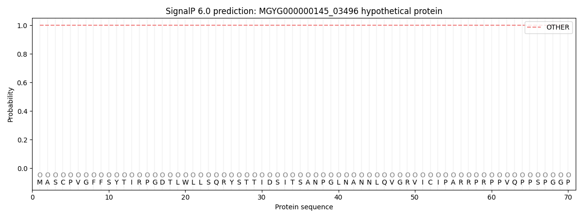
This protein is predicted as OTHER
| Other | SP_Sec_SPI | LIPO_Sec_SPII | TAT_Tat_SPI | TATLIP_Sec_SPII | PILIN_Sec_SPIII |
|---|---|---|---|---|---|
| 1.000078 | 0.000001 | 0.000000 | 0.000000 | 0.000000 | 0.000000 |