You are browsing environment: HUMAN GUT
CAZyme Information: MGYG000000147_01656
You are here: Home > Sequence: MGYG000000147_01656
Basic Information |
Genomic context |
Full Sequence |
Enzyme annotations |
CAZy signature domains |
CDD domains |
CAZyme hits |
PDB hits |
Swiss-Prot hits |
SignalP and Lipop annotations |
TMHMM annotations
Basic Information help
| Species | Bacillus paralicheniformis | |||||||||||
|---|---|---|---|---|---|---|---|---|---|---|---|---|
| Lineage | Bacteria; Firmicutes; Bacilli; Bacillales; Bacillaceae; Bacillus; Bacillus paralicheniformis | |||||||||||
| CAZyme ID | MGYG000000147_01656 | |||||||||||
| CAZy Family | CBM50 | |||||||||||
| CAZyme Description | N-acetylmuramoyl-L-alanine amidase CwlA | |||||||||||
| CAZyme Property |
|
|||||||||||
| Genome Property |
|
|||||||||||
| Gene Location | Start: 737338; End: 738330 Strand: + | |||||||||||
CDD Domains download full data without filtering help
| Cdd ID | Domain | E-Value | qStart | qEnd | sStart | sEnd | Domain Description |
|---|---|---|---|---|---|---|---|
| COG5632 | CwlA | 8.27e-59 | 3 | 161 | 2 | 170 | N-acetylmuramoyl-L-alanine amidase CwlA [Cell wall/membrane/envelope biogenesis]. |
| cd06583 | PGRP | 6.47e-30 | 24 | 142 | 1 | 125 | Peptidoglycan recognition proteins (PGRPs) are pattern recognition receptors that bind, and in certain cases, hydrolyze peptidoglycans (PGNs) of bacterial cell walls. PGRPs have been divided into three classes: short PGRPs (PGRP-S), that are small (20 kDa) extracellular proteins; intermediate PGRPs (PGRP-I) that are 40-45 kDa and are predicted to be transmembrane proteins; and long PGRPs (PGRP-L), up to 90 kDa, which may be either intracellular or transmembrane. Several structures of PGRPs are known in insects and mammals, some bound with substrates like Muramyl Tripeptide (MTP) or Tracheal Cytotoxin (TCT). The substrate binding site is conserved in PGRP-LCx, PGRP-LE, and PGRP-Ialpha proteins. This family includes Zn-dependent N-Acetylmuramoyl-L-alanine Amidase, EC:3.5.1.28. This enzyme cleaves the amide bond between N-acetylmuramoyl and L-amino acids, preferentially D-lactyl-L-Ala, in bacterial cell walls. The structure for the bacteriophage T7 lysozyme shows that two of the conserved histidines and a cysteine are zinc binding residues. Site-directed mutagenesis of T7 lysozyme indicates that two conserved residues, a Tyr and a Lys, are important for amidase activity. |
| smart00644 | Ami_2 | 7.41e-29 | 23 | 140 | 1 | 126 | Ami_2 domain. |
| pfam01510 | Amidase_2 | 2.17e-27 | 26 | 142 | 3 | 121 | N-acetylmuramoyl-L-alanine amidase. This family includes zinc amidases that have N-acetylmuramoyl-L-alanine amidase activity EC:3.5.1.28. This enzyme domain cleaves the amide bond between N-acetylmuramoyl and L-amino acids in bacterial cell walls (preferentially: D-lactyl-L-Ala). The structure is known for the bacteriophage T7 structure and shows that two of the conserved histidines are zinc binding. |
| cd00118 | LysM | 2.94e-17 | 178 | 221 | 2 | 45 | Lysin Motif is a small domain involved in binding peptidoglycan. LysM, a small globular domain with approximately 40 amino acids, is a widespread protein module involved in binding peptidoglycan in bacteria and chitin in eukaryotes. The domain was originally identified in enzymes that degrade bacterial cell walls, but proteins involved in many other biological functions also contain this domain. It has been reported that the LysM domain functions as a signal for specific plant-bacteria recognition in bacterial pathogenesis. Many of these enzymes are modular and are composed of catalytic units linked to one or several repeats of LysM domains. LysM domains are found in bacteria and eukaryotes. |
CAZyme Hits help
| Hit ID | E-Value | Query Start | Query End | Hit Start | Hit End |
|---|---|---|---|---|---|
| QSV41397.1 | 1.49e-226 | 1 | 330 | 1 | 330 |
| QBJ82979.1 | 4.94e-166 | 1 | 323 | 1 | 324 |
| QHL55113.1 | 4.94e-166 | 1 | 323 | 1 | 324 |
| ADM37946.1 | 1.62e-162 | 1 | 323 | 1 | 325 |
| QDD03075.1 | 1.62e-162 | 1 | 323 | 1 | 325 |
PDB Hits download full data without filtering help
| Hit ID | E-Value | Query Start | Query End | Hit Start | Hit End | Description |
|---|---|---|---|---|---|---|
| 1YB0_A | 9.69e-64 | 3 | 151 | 1 | 149 | Structureof PlyL [Bacillus anthracis],1YB0_B Structure of PlyL [Bacillus anthracis],1YB0_C Structure of PlyL [Bacillus anthracis] |
| 2AR3_A | 8.03e-63 | 3 | 151 | 1 | 149 | ChainA, prophage lambdaba02, n-acetylmuramoyl-l-alanine amidase, family 2 [Bacillus anthracis],2AR3_B Chain B, prophage lambdaba02, n-acetylmuramoyl-l-alanine amidase, family 2 [Bacillus anthracis],2AR3_C Chain C, prophage lambdaba02, n-acetylmuramoyl-l-alanine amidase, family 2 [Bacillus anthracis] |
| 2L47_A | 5.35e-62 | 3 | 151 | 1 | 149 | Solutionstructure of the PlyG catalytic domain [Bacillus phage Gamma] |
| 3HMB_A | 4.04e-32 | 21 | 156 | 22 | 156 | ChainA, N-acetylmuramoyl-L-alanine amidase xlyA [Bacillus subtilis],3HMB_B Chain B, N-acetylmuramoyl-L-alanine amidase xlyA [Bacillus subtilis],3HMB_C Chain C, N-acetylmuramoyl-L-alanine amidase xlyA [Bacillus subtilis] |
| 3RDR_A | 8.93e-30 | 21 | 156 | 22 | 156 | Structureof the catalytic domain of XlyA [Bacillus subtilis] |
Swiss-Prot Hits download full data without filtering help
| Hit ID | E-Value | Query Start | Query End | Hit Start | Hit End | Description |
|---|---|---|---|---|---|---|
| O34391 | 6.12e-145 | 1 | 322 | 1 | 317 | N-acetylmuramoyl-L-alanine amidase XlyB OS=Bacillus subtilis (strain 168) OX=224308 GN=xlyB PE=3 SV=1 |
| P24808 | 1.02e-125 | 1 | 323 | 1 | 272 | N-acetylmuramoyl-L-alanine amidase CwlA OS=Bacillus subtilis (strain 168) OX=224308 GN=cwlA PE=1 SV=1 |
| P39800 | 6.40e-80 | 21 | 322 | 19 | 296 | N-acetylmuramoyl-L-alanine amidase XlyA OS=Bacillus subtilis (strain 168) OX=224308 GN=xlyA PE=1 SV=1 |
| P54450 | 1.17e-53 | 3 | 323 | 2 | 250 | N-acetylmuramoyl-L-alanine amidase CwlH OS=Bacillus subtilis (strain 168) OX=224308 GN=cwlH PE=1 SV=1 |
| P36550 | 2.54e-51 | 3 | 323 | 2 | 360 | N-acetylmuramoyl-L-alanine amidase CwlL OS=Bacillus licheniformis OX=1402 GN=cwlL PE=3 SV=1 |
SignalP and Lipop Annotations help
This protein is predicted as OTHER
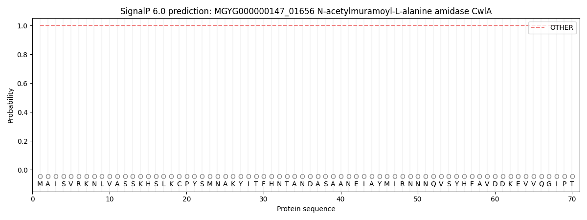
| Other | SP_Sec_SPI | LIPO_Sec_SPII | TAT_Tat_SPI | TATLIP_Sec_SPII | PILIN_Sec_SPIII |
|---|---|---|---|---|---|
| 1.000049 | 0.000000 | 0.000000 | 0.000000 | 0.000000 | 0.000000 |