You are browsing environment: HUMAN GUT
CAZyme Information: MGYG000000162_00642
You are here: Home > Sequence: MGYG000000162_00642
Basic Information |
Genomic context |
Full Sequence |
Enzyme annotations |
CAZy signature domains |
CDD domains |
CAZyme hits |
PDB hits |
Swiss-Prot hits |
SignalP and Lipop annotations |
TMHMM annotations
Basic Information help
| Species | Enterococcus_B durans | |||||||||||
|---|---|---|---|---|---|---|---|---|---|---|---|---|
| Lineage | Bacteria; Firmicutes; Bacilli; Lactobacillales; Enterococcaceae; Enterococcus_B; Enterococcus_B durans | |||||||||||
| CAZyme ID | MGYG000000162_00642 | |||||||||||
| CAZy Family | GH13 | |||||||||||
| CAZyme Description | Trehalose-6-phosphate hydrolase | |||||||||||
| CAZyme Property |
|
|||||||||||
| Genome Property |
|
|||||||||||
| Gene Location | Start: 4557; End: 5582 Strand: - | |||||||||||
CAZyme Signature Domains help
| Family | Start | End | Evalue | family coverage |
|---|---|---|---|---|
| GH13 | 24 | 340 | 3.4e-155 | 0.9302325581395349 |
CDD Domains download full data without filtering help
| Cdd ID | Domain | E-Value | qStart | qEnd | sStart | sEnd | Domain Description |
|---|---|---|---|---|---|---|---|
| TIGR02403 | trehalose_treC | 0.0 | 3 | 340 | 2 | 342 | alpha,alpha-phosphotrehalase. Trehalose is a glucose disaccharide that serves in many biological systems as a compatible solute for protection against hyperosmotic and thermal stress. This family describes trehalose-6-phosphate hydrolase, product of the treC (or treA) gene, which is often found together with a trehalose uptake transporter and a trehalose operon repressor. |
| cd11333 | AmyAc_SI_OligoGlu_DGase | 0.0 | 4 | 340 | 1 | 346 | Alpha amylase catalytic domain found in Sucrose isomerases, oligo-1,6-glucosidase (also called isomaltase; sucrase-isomaltase; alpha-limit dextrinase), dextran glucosidase (also called glucan 1,6-alpha-glucosidase), and related proteins. The sucrose isomerases (SIs) Isomaltulose synthase (EC 5.4.99.11) and Trehalose synthase (EC 5.4.99.16) catalyze the isomerization of sucrose and maltose to produce isomaltulose and trehalulose, respectively. Oligo-1,6-glucosidase (EC 3.2.1.10) hydrolyzes the alpha-1,6-glucosidic linkage of isomaltooligosaccharides, pannose, and dextran. Unlike alpha-1,4-glucosidases (EC 3.2.1.20), it fails to hydrolyze the alpha-1,4-glucosidic bonds of maltosaccharides. Dextran glucosidase (DGase, EC 3.2.1.70) hydrolyzes alpha-1,6-glucosidic linkages at the non-reducing end of panose, isomaltooligosaccharides and dextran to produce alpha-glucose.The common reaction chemistry of the alpha-amylase family enzymes is based on a two-step acid catalytic mechanism that requires two critical carboxylates: one acting as a general acid/base (Glu) and the other as a nucleophile (Asp). Both hydrolysis and transglycosylation proceed via the nucleophilic substitution reaction between the anomeric carbon, C1 and a nucleophile. Both enzymes contain the three catalytic residues (Asp, Glu and Asp) common to the alpha-amylase family as well as two histidine residues which are predicted to be critical to binding the glucose residue adjacent to the scissile bond in the substrates. The Alpha-amylase family comprises the largest family of glycoside hydrolases (GH), with the majority of enzymes acting on starch, glycogen, and related oligo- and polysaccharides. These proteins catalyze the transformation of alpha-1,4 and alpha-1,6 glucosidic linkages with retention of the anomeric center. The protein is described as having 3 domains: A, B, C. A is a (beta/alpha) 8-barrel; B is a loop between the beta 3 strand and alpha 3 helix of A; C is the C-terminal extension characterized by a Greek key. The majority of the enzymes have an active site cleft found between domains A and B where a triad of catalytic residues performs catalysis. Other members of this family have lost the catalytic activity as in the case of the human 4F2hc, or only have 2 residues that serve as the catalytic nucleophile and the acid/base, such as Thermus A4 beta-galactosidase with 2 Glu residues (GH42) and human alpha-galactosidase with 2 Asp residues (GH31). The family members are quite extensive and include: alpha amylase, maltosyltransferase, cyclodextrin glycotransferase, maltogenic amylase, neopullulanase, isoamylase, 1,4-alpha-D-glucan maltotetrahydrolase, 4-alpha-glucotransferase, oligo-1,6-glucosidase, amylosucrase, sucrose phosphorylase, and amylomaltase. |
| PRK10933 | PRK10933 | 2.80e-155 | 7 | 340 | 12 | 348 | trehalose-6-phosphate hydrolase; Provisional |
| pfam00128 | Alpha-amylase | 3.14e-109 | 25 | 325 | 1 | 297 | Alpha amylase, catalytic domain. Alpha amylase is classified as family 13 of the glycosyl hydrolases. The structure is an 8 stranded alpha/beta barrel containing the active site, interrupted by a ~70 a.a. calcium-binding domain protruding between beta strand 3 and alpha helix 3, and a carboxyl-terminal Greek key beta-barrel domain. |
| cd11331 | AmyAc_OligoGlu_like | 5.42e-105 | 4 | 325 | 4 | 330 | Alpha amylase catalytic domain found in oligo-1,6-glucosidase (also called isomaltase; sucrase-isomaltase; alpha-limit dextrinase) and related proteins. Oligo-1,6-glucosidase (EC 3.2.1.10) hydrolyzes the alpha-1,6-glucosidic linkage of isomalto-oligosaccharides, pannose, and dextran. Unlike alpha-1,4-glucosidases (EC 3.2.1.20), it fails to hydrolyze the alpha-1,4-glucosidic bonds of maltosaccharides. The Alpha-amylase family comprises the largest family of glycoside hydrolases (GH), with the majority of enzymes acting on starch, glycogen, and related oligo- and polysaccharides. These proteins catalyze the transformation of alpha-1,4 and alpha-1,6 glucosidic linkages with retention of the anomeric center. The protein is described as having 3 domains: A, B, C. A is a (beta/alpha) 8-barrel; B is a loop between the beta 3 strand and alpha 3 helix of A; C is the C-terminal extension characterized by a Greek key. The majority of the enzymes have an active site cleft found between domains A and B where a triad of catalytic residues (Asp, Glu and Asp) performs catalysis. Other members of this family have lost the catalytic activity as in the case of the human 4F2hc, or only have 2 residues that serve as the catalytic nucleophile and the acid/base, such as Thermus A4 beta-galactosidase with 2 Glu residues (GH42) and human alpha-galactosidase with 2 Asp residues (GH31). The family members are quite extensive and include: alpha amylase, maltosyltransferase, cyclodextrin glycotransferase, maltogenic amylase, neopullulanase, isoamylase, 1,4-alpha-D-glucan maltotetrahydrolase, 4-alpha-glucotransferase, oligo-1,6-glucosidase, amylosucrase, sucrose phosphorylase, and amylomaltase. |
CAZyme Hits help
| Hit ID | E-Value | Query Start | Query End | Hit Start | Hit End |
|---|---|---|---|---|---|
| ASV94691.1 | 5.79e-262 | 1 | 340 | 1 | 340 |
| AKX85571.1 | 1.93e-260 | 1 | 340 | 1 | 340 |
| AKZ49222.1 | 1.93e-260 | 1 | 340 | 1 | 340 |
| QED58858.1 | 7.83e-260 | 1 | 340 | 1 | 340 |
| QPQ26994.1 | 4.52e-259 | 1 | 340 | 1 | 340 |
PDB Hits download full data without filtering help
| Hit ID | E-Value | Query Start | Query End | Hit Start | Hit End | Description |
|---|---|---|---|---|---|---|
| 5BRQ_A | 8.92e-135 | 3 | 340 | 15 | 358 | Crystalstructure of Bacillus licheniformis trehalose-6-phosphate hydrolase (TreA) [Bacillus licheniformis DSM 13 = ATCC 14580],5BRQ_B Crystal structure of Bacillus licheniformis trehalose-6-phosphate hydrolase (TreA) [Bacillus licheniformis DSM 13 = ATCC 14580],5BRQ_C Crystal structure of Bacillus licheniformis trehalose-6-phosphate hydrolase (TreA) [Bacillus licheniformis DSM 13 = ATCC 14580],5BRQ_D Crystal structure of Bacillus licheniformis trehalose-6-phosphate hydrolase (TreA) [Bacillus licheniformis DSM 13 = ATCC 14580] |
| 5BRP_A | 3.58e-134 | 3 | 340 | 15 | 358 | Crystalstructure of Bacillus licheniformis trehalose-6-phosphate hydrolase (TreA), mutant R201Q, in complex with PNG [Bacillus licheniformis DSM 13 = ATCC 14580],5BRP_B Crystal structure of Bacillus licheniformis trehalose-6-phosphate hydrolase (TreA), mutant R201Q, in complex with PNG [Bacillus licheniformis DSM 13 = ATCC 14580],5BRP_C Crystal structure of Bacillus licheniformis trehalose-6-phosphate hydrolase (TreA), mutant R201Q, in complex with PNG [Bacillus licheniformis DSM 13 = ATCC 14580],5BRP_D Crystal structure of Bacillus licheniformis trehalose-6-phosphate hydrolase (TreA), mutant R201Q, in complex with PNG [Bacillus licheniformis DSM 13 = ATCC 14580] |
| 4AIE_A | 8.06e-106 | 3 | 340 | 7 | 341 | Structureof glucan-1,6-alpha-glucosidase from Lactobacillus acidophilus NCFM [Lactobacillus acidophilus NCFM] |
| 1UOK_A | 1.16e-104 | 3 | 340 | 6 | 352 | CrystalStructure Of B. Cereus Oligo-1,6-Glucosidase [Bacillus cereus] |
| 5DO8_A | 1.88e-99 | 3 | 340 | 7 | 351 | 1.8Angstrom crystal structure of Listeria monocytogenes Lmo0184 alpha-1,6-glucosidase [Listeria monocytogenes EGD-e],5DO8_B 1.8 Angstrom crystal structure of Listeria monocytogenes Lmo0184 alpha-1,6-glucosidase [Listeria monocytogenes EGD-e],5DO8_C 1.8 Angstrom crystal structure of Listeria monocytogenes Lmo0184 alpha-1,6-glucosidase [Listeria monocytogenes EGD-e] |
Swiss-Prot Hits download full data without filtering help
| Hit ID | E-Value | Query Start | Query End | Hit Start | Hit End | Description |
|---|---|---|---|---|---|---|
| P39795 | 1.65e-130 | 3 | 340 | 9 | 352 | Trehalose-6-phosphate hydrolase OS=Bacillus subtilis (strain 168) OX=224308 GN=treA PE=1 SV=2 |
| P28904 | 2.17e-120 | 3 | 340 | 8 | 348 | Trehalose-6-phosphate hydrolase OS=Escherichia coli (strain K12) OX=83333 GN=treC PE=1 SV=3 |
| P29094 | 3.99e-109 | 3 | 340 | 6 | 353 | Oligo-1,6-glucosidase OS=Parageobacillus thermoglucosidasius OX=1426 GN=malL PE=1 SV=1 |
| Q54796 | 2.97e-105 | 7 | 340 | 10 | 336 | Glucan 1,6-alpha-glucosidase OS=Streptococcus pneumoniae serotype 4 (strain ATCC BAA-334 / TIGR4) OX=170187 GN=dexB PE=3 SV=2 |
| P21332 | 6.37e-104 | 3 | 340 | 6 | 352 | Oligo-1,6-glucosidase OS=Bacillus cereus OX=1396 GN=malL PE=1 SV=1 |
SignalP and Lipop Annotations help
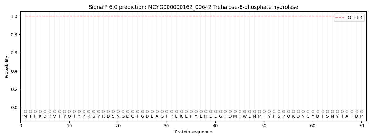
This protein is predicted as OTHER

| Other | SP_Sec_SPI | LIPO_Sec_SPII | TAT_Tat_SPI | TATLIP_Sec_SPII | PILIN_Sec_SPIII |
|---|---|---|---|---|---|
| 1.000076 | 0.000001 | 0.000000 | 0.000000 | 0.000000 | 0.000000 |
