You are browsing environment: HUMAN GUT
CAZyme Information: MGYG000000174_01940
You are here: Home > Sequence: MGYG000000174_01940
Basic Information |
Genomic context |
Full Sequence |
Enzyme annotations |
CAZy signature domains |
CDD domains |
CAZyme hits |
PDB hits |
Swiss-Prot hits |
SignalP and Lipop annotations |
TMHMM annotations
Basic Information help
| Species | Parabacteroides faecis | |||||||||||
|---|---|---|---|---|---|---|---|---|---|---|---|---|
| Lineage | Bacteria; Bacteroidota; Bacteroidia; Bacteroidales; Tannerellaceae; Parabacteroides; Parabacteroides faecis | |||||||||||
| CAZyme ID | MGYG000000174_01940 | |||||||||||
| CAZy Family | CBM50 | |||||||||||
| CAZyme Description | hypothetical protein | |||||||||||
| CAZyme Property |
|
|||||||||||
| Genome Property |
|
|||||||||||
| Gene Location | Start: 221621; End: 223420 Strand: - | |||||||||||
CDD Domains download full data without filtering help
| Cdd ID | Domain | E-Value | qStart | qEnd | sStart | sEnd | Domain Description |
|---|---|---|---|---|---|---|---|
| cd00118 | LysM | 7.05e-10 | 41 | 83 | 2 | 45 | Lysin Motif is a small domain involved in binding peptidoglycan. LysM, a small globular domain with approximately 40 amino acids, is a widespread protein module involved in binding peptidoglycan in bacteria and chitin in eukaryotes. The domain was originally identified in enzymes that degrade bacterial cell walls, but proteins involved in many other biological functions also contain this domain. It has been reported that the LysM domain functions as a signal for specific plant-bacteria recognition in bacterial pathogenesis. Many of these enzymes are modular and are composed of catalytic units linked to one or several repeats of LysM domains. LysM domains are found in bacteria and eukaryotes. |
| pfam01476 | LysM | 1.52e-07 | 42 | 84 | 1 | 43 | LysM domain. The LysM (lysin motif) domain is about 40 residues long. It is found in a variety of enzymes involved in bacterial cell wall degradation. This domain may have a general peptidoglycan binding function. The structure of this domain is known. |
| smart00257 | LysM | 5.42e-07 | 41 | 83 | 1 | 44 | Lysin motif. |
| cd06347 | PBP1_ABC_LivK_ligand_binding-like | 7.18e-07 | 271 | 548 | 21 | 295 | type 1 periplasmic ligand-binding domain of uncharacterized ABC (Atpase Binding Cassette)-type active transport systems predicted to be involved in uptake of amino acids, peptides, or inorganic ions. This subgroup includes the type 1 periplasmic ligand-binding domain of uncharacterized ABC (Atpase Binding Cassette)-type active transport systems that are predicted to be involved in uptake of amino acids, peptides, or inorganic ions. This subgroup has high sequence similarity to members of the family of hydrophobic amino acid transporters (HAAT), such as leucine-isoleucine-valine binding protein (LIVBP); however, its ligand specificity has not been determined experimentally. |
| smart00257 | LysM | 1.45e-06 | 102 | 145 | 1 | 44 | Lysin motif. |
CAZyme Hits help
| Hit ID | E-Value | Query Start | Query End | Hit Start | Hit End |
|---|---|---|---|---|---|
| QUT48586.1 | 0.0 | 1 | 599 | 1 | 600 |
| QUR46882.1 | 3.20e-316 | 1 | 599 | 1 | 602 |
| QJE27971.1 | 3.20e-316 | 1 | 599 | 1 | 602 |
| QCY56025.1 | 3.20e-316 | 1 | 599 | 1 | 602 |
| ABR42587.1 | 3.20e-316 | 1 | 599 | 1 | 602 |
Swiss-Prot Hits download full data without filtering help
| Hit ID | E-Value | Query Start | Query End | Hit Start | Hit End | Description |
|---|---|---|---|---|---|---|
| O05495 | 5.49e-06 | 39 | 147 | 1 | 94 | Putative sporulation-specific glycosylase YdhD OS=Bacillus subtilis (strain 168) OX=224308 GN=ydhD PE=1 SV=2 |
SignalP and Lipop Annotations help
This protein is predicted as SP
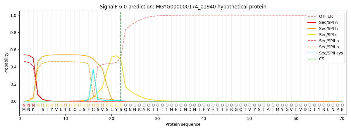
| Other | SP_Sec_SPI | LIPO_Sec_SPII | TAT_Tat_SPI | TATLIP_Sec_SPII | PILIN_Sec_SPIII |
|---|---|---|---|---|---|
| 0.001281 | 0.527152 | 0.470661 | 0.000330 | 0.000290 | 0.000258 |