You are browsing environment: HUMAN GUT
CAZyme Information: MGYG000000191_01693
You are here: Home > Sequence: MGYG000000191_01693
Basic Information |
Genomic context |
Full Sequence |
Enzyme annotations |
CAZy signature domains |
CDD domains |
CAZyme hits |
PDB hits |
Swiss-Prot hits |
SignalP and Lipop annotations |
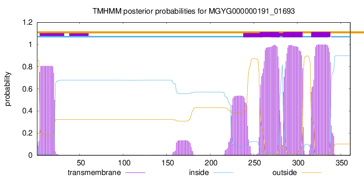
TMHMM annotations
Basic Information help
| Species | Weizmannia coagulans | |||||||||||
|---|---|---|---|---|---|---|---|---|---|---|---|---|
| Lineage | Bacteria; Firmicutes; Bacilli; Bacillales_B; Bacillaceae_C; Weizmannia; Weizmannia coagulans | |||||||||||
| CAZyme ID | MGYG000000191_01693 | |||||||||||
| CAZy Family | GT2 | |||||||||||
| CAZyme Description | 4,4'-diaponeurosporenoate glycosyltransferase | |||||||||||
| CAZyme Property |
|
|||||||||||
| Genome Property |
|
|||||||||||
| Gene Location | Start: 5618; End: 6703 Strand: - | |||||||||||
CAZyme Signature Domains help
| Family | Start | End | Evalue | family coverage |
|---|---|---|---|---|
| GT2 | 38 | 160 | 6.3e-23 | 0.7352941176470589 |
CDD Domains download full data without filtering help
| Cdd ID | Domain | E-Value | qStart | qEnd | sStart | sEnd | Domain Description |
|---|---|---|---|---|---|---|---|
| cd00761 | Glyco_tranf_GTA_type | 1.84e-22 | 39 | 150 | 1 | 113 | Glycosyltransferase family A (GT-A) includes diverse families of glycosyl transferases with a common GT-A type structural fold. Glycosyltransferases (GTs) are enzymes that synthesize oligosaccharides, polysaccharides, and glycoconjugates by transferring the sugar moiety from an activated nucleotide-sugar donor to an acceptor molecule, which may be a growing oligosaccharide, a lipid, or a protein. Based on the stereochemistry of the donor and acceptor molecules, GTs are classified as either retaining or inverting enzymes. To date, all GT structures adopt one of two possible folds, termed GT-A fold and GT-B fold. This hierarchy includes diverse families of glycosyl transferases with a common GT-A type structural fold, which has two tightly associated beta/alpha/beta domains that tend to form a continuous central sheet of at least eight beta-strands. The majority of the proteins in this superfamily are Glycosyltransferase family 2 (GT-2) proteins. But it also includes families GT-43, GT-6, GT-8, GT13 and GT-7; which are evolutionarily related to GT-2 and share structure similarities. |
| cd02522 | GT_2_like_a | 4.17e-22 | 37 | 229 | 1 | 177 | GT_2_like_a represents a glycosyltransferase family-2 subfamily with unknown function. Glycosyltransferase family 2 (GT-2) subfamily of unknown function. GT-2 includes diverse families of glycosyltransferases with a common GT-A type structural fold, which has two tightly associated beta/alpha/beta domains that tend to form a continuous central sheet of at least eight beta-strands. These are enzymes that catalyze the transfer of sugar moieties from activated donor molecules to specific acceptor molecules, forming glycosidic bonds. Glycosyltransferases have been classified into more than 90 distinct sequence based families. |
| cd06423 | CESA_like | 1.98e-21 | 39 | 203 | 1 | 171 | CESA_like is the cellulose synthase superfamily. The cellulose synthase (CESA) superfamily includes a wide variety of glycosyltransferase family 2 enzymes that share the common characteristic of catalyzing the elongation of polysaccharide chains. The members include cellulose synthase catalytic subunit, chitin synthase, glucan biosynthesis protein and other families of CESA-like proteins. Cellulose synthase catalyzes the polymerization reaction of cellulose, an aggregate of unbranched polymers of beta-1,4-linked glucose residues in plants, most algae, some bacteria and fungi, and even some animals. In bacteria, algae and lower eukaryotes, there is a second unrelated type of cellulose synthase (Type II), which produces acylated cellulose, a derivative of cellulose. Chitin synthase catalyzes the incorporation of GlcNAc from substrate UDP-GlcNAc into chitin, which is a linear homopolymer of beta-(1,4)-linked GlcNAc residues and Glucan Biosynthesis protein catalyzes the elongation of beta-1,2 polyglucose chains of Glucan. |
| pfam00535 | Glycos_transf_2 | 6.85e-21 | 38 | 150 | 1 | 113 | Glycosyl transferase family 2. Diverse family, transferring sugar from UDP-glucose, UDP-N-acetyl- galactosamine, GDP-mannose or CDP-abequose, to a range of substrates including cellulose, dolichol phosphate and teichoic acids. |
| COG0463 | WcaA | 1.67e-20 | 36 | 341 | 4 | 291 | Glycosyltransferase involved in cell wall bisynthesis [Cell wall/membrane/envelope biogenesis]. |
CAZyme Hits help
| Hit ID | E-Value | Query Start | Query End | Hit Start | Hit End |
|---|---|---|---|---|---|
| AEH52282.1 | 1.65e-266 | 1 | 361 | 1 | 361 |
| QJE31752.1 | 3.19e-264 | 1 | 361 | 1 | 361 |
| AJH78691.1 | 3.19e-264 | 1 | 361 | 1 | 361 |
| QWU06847.1 | 8.28e-251 | 1 | 361 | 1 | 361 |
| AKN53887.1 | 7.93e-249 | 1 | 361 | 1 | 361 |
PDB Hits download full data without filtering help
| Hit ID | E-Value | Query Start | Query End | Hit Start | Hit End | Description |
|---|---|---|---|---|---|---|
| 3F1Y_A | 1.26e-09 | 37 | 126 | 96 | 191 | Mannosyl-3-phosphoglyceratesynthase from Rubrobacter xylanophilus [synthetic construct],3F1Y_C Mannosyl-3-phosphoglycerate synthase from Rubrobacter xylanophilus [synthetic construct],3KIA_A Crystal structure of mannosyl-3-phosphoglycerate synthase from Rubrobacter xylanophilus [synthetic construct],3KIA_C Crystal structure of mannosyl-3-phosphoglycerate synthase from Rubrobacter xylanophilus [synthetic construct],3O3P_A Crystal structure of R. xylanophilus MpgS in complex with GDP-Mannose [Rubrobacter xylanophilus],3O3P_B Crystal structure of R. xylanophilus MpgS in complex with GDP-Mannose [Rubrobacter xylanophilus] |
| 4Y6N_A | 2.48e-07 | 32 | 124 | 45 | 140 | Crystalstructure of glucosyl-3-phosphoglycerate synthase from Mycobacterium tuberculosis in complex with Mn2+, uridine-diphosphate-glucose (UDP-Glc) and phosphoglyceric acid (PGA) - GpgS Mn2+ UDP-Glc PGA-1 [Mycobacterium tuberculosis H37Rv],4Y6U_A Mycobacterial protein [Mycobacterium tuberculosis H37Rv],4Y7F_A Crystal structure of glucosyl-3-phosphoglycerate synthase from Mycobacterium tuberculosis in complex with Mn2+, uridine-diphosphate-glucose (UDP-Glc) and 3-(phosphonooxy)propanoic acid (PPA) - GpgS Mn2+ UDP-Glc PPA [Mycobacterium tuberculosis H37Rv],4Y7G_A Crystal structure of glucosyl-3-phosphoglycerate synthase from Mycobacterium tuberculosis in complex with Mn2+, uridine-diphosphate-glucose (UDP-Glc) and glycerol 3-phosphate (G3P) - GpgS Mn2+ UDP-Glc G3P [Mycobacterium tuberculosis H37Rv],4Y9X_A Crystal structure of glucosyl-3-phosphoglycerate synthase from Mycobacterium tuberculosis in complex with Mn2+, uridine-diphosphate-glucose (UDP-Glc) and phosphoglyceric acid (PGA) - GpgS Mn2+ UDP-Glc PGA-3 [Mycobacterium tuberculosis H37Rv],5JQX_A Crystal structure of glucosyl-3-phosphoglycerate synthase from Mycobacterium tuberculosis in complex with phosphoglyceric acid (PGA) - GpgS*PGA [Mycobacterium tuberculosis H37Ra],5JQX_B Crystal structure of glucosyl-3-phosphoglycerate synthase from Mycobacterium tuberculosis in complex with phosphoglyceric acid (PGA) - GpgS*PGA [Mycobacterium tuberculosis H37Ra],5JQX_C Crystal structure of glucosyl-3-phosphoglycerate synthase from Mycobacterium tuberculosis in complex with phosphoglyceric acid (PGA) - GpgS*PGA [Mycobacterium tuberculosis H37Ra],5JQX_D Crystal structure of glucosyl-3-phosphoglycerate synthase from Mycobacterium tuberculosis in complex with phosphoglyceric acid (PGA) - GpgS*PGA [Mycobacterium tuberculosis H37Ra],5JSX_A Crystal structure of glucosyl-3-phosphoglycerate synthase from Mycobacterium tuberculosis in complex with Mn2+ and uridine-diphosphate-glucose (UDP-Glc) [Mycobacterium tuberculosis H37Ra],5JT0_A Crystal structure of glucosyl-3-phosphoglycerate synthase from Mycobacterium tuberculosis in complex with Mn2+, uridine-diphosphate (UDP) and glucosyl-3-phosphoglycerate (GPG) - GpgS*GPG*UDP*Mn2+ [Mycobacterium tuberculosis H37Rv],5JUC_A Crystal structure of glucosyl-3-phosphoglycerate synthase from Mycobacterium tuberculosis in complex with Mn2+, uridine-diphosphate (UDP) and glucosyl-3-phosphoglycerate (GPG) - GpgS*GPG*UDP*Mn2+_2 [Mycobacterium tuberculosis H37Rv],5JUD_A Crystal structure of glucosyl-3-phosphoglycerate synthase from Mycobacterium tuberculosis in complex with uridine-diphosphate (UDP) - GpgS*UDP [Mycobacterium tuberculosis variant bovis AF2122/97] |
| 3E25_A | 2.56e-07 | 32 | 124 | 41 | 136 | ChainA, Crystal structure of M. tuberculosis glucosyl-3-phosphoglycerate synthase [Mycobacterium tuberculosis],3E26_A Chain A, Crystal structure of M. tuberculosis glucosyl-3-phosphoglycerate synthase [Mycobacterium tuberculosis] |
| 4DDZ_A | 2.62e-07 | 32 | 124 | 61 | 156 | Crystalstructure of glucosyl-3-phosphoglycerate synthase from Mycobacterium tuberculosis [Mycobacterium tuberculosis H37Rv],4DE7_A Crystal structure of glucosyl-3-phosphoglycerate synthase from Mycobacterium tuberculosis in complex with Mg2+ and uridine-diphosphate (UDP) [Mycobacterium tuberculosis H37Rv],4DEC_A Crystal structure of glucosyl-3-phosphoglycerate synthase from Mycobacterium tuberculosis in complex with Mn2+, uridine-diphosphate (UDP) and phosphoglyceric acid (PGA) [Mycobacterium tuberculosis H37Rv],5JQQ_A Crystal structure of glucosyl-3-phosphoglycerate synthase from Mycobacterium tuberculosis - apo form [Mycobacterium tuberculosis H37Ra] |
| 3CKJ_A | 4.43e-07 | 32 | 124 | 46 | 141 | CrystalStructure of a Mycobacterial Protein [Mycobacterium avium subsp. paratuberculosis],3CKN_A Crystal Structure of a Mycobacterial Protein [Mycobacterium avium subsp. paratuberculosis],3CKO_A Crystal Structure of a Mycobacterial Protein [Mycobacterium avium subsp. paratuberculosis],3CKQ_A Crystal Structure of a Mycobacterial Protein [Mycobacterium avium subsp. paratuberculosis],3CKV_A Crystal Structure of a Mycobacterial Protein [Mycobacterium avium subsp. paratuberculosis] |
Swiss-Prot Hits download full data without filtering help
| Hit ID | E-Value | Query Start | Query End | Hit Start | Hit End | Description |
|---|---|---|---|---|---|---|
| Q2FDU4 | 3.59e-54 | 9 | 359 | 16 | 375 | 4,4'-diaponeurosporenoate glycosyltransferase OS=Staphylococcus aureus (strain USA300) OX=367830 GN=crtQ PE=3 SV=1 |
| Q6G6B1 | 3.59e-54 | 9 | 359 | 16 | 375 | 4,4'-diaponeurosporenoate glycosyltransferase OS=Staphylococcus aureus (strain MSSA476) OX=282459 GN=crtQ PE=3 SV=1 |
| Q53590 | 3.59e-54 | 9 | 359 | 16 | 375 | 4,4'-diaponeurosporenoate glycosyltransferase OS=Staphylococcus aureus (strain Newman) OX=426430 GN=crtQ PE=3 SV=2 |
| Q5HCY7 | 3.59e-54 | 9 | 359 | 16 | 375 | 4,4'-diaponeurosporenoate glycosyltransferase OS=Staphylococcus aureus (strain COL) OX=93062 GN=crtQ PE=3 SV=2 |
| Q2FV58 | 3.59e-54 | 9 | 359 | 16 | 375 | 4,4'-diaponeurosporenoate glycosyltransferase OS=Staphylococcus aureus (strain NCTC 8325 / PS 47) OX=93061 GN=crtQ PE=3 SV=1 |
SignalP and Lipop Annotations help
This protein is predicted as OTHER

| Other | SP_Sec_SPI | LIPO_Sec_SPII | TAT_Tat_SPI | TATLIP_Sec_SPII | PILIN_Sec_SPIII |
|---|---|---|---|---|---|
| 1.000045 | 0.000003 | 0.000000 | 0.000000 | 0.000000 | 0.000000 |