You are browsing environment: HUMAN GUT
CAZyme Information: MGYG000000198_01945
You are here: Home > Sequence: MGYG000000198_01945
Basic Information |
Genomic context |
Full Sequence |
Enzyme annotations |
CAZy signature domains |
CDD domains |
CAZyme hits |
PDB hits |
Swiss-Prot hits |
SignalP and Lipop annotations |
TMHMM annotations
Basic Information help
| Species | Enterocloster citroniae | |||||||||||
|---|---|---|---|---|---|---|---|---|---|---|---|---|
| Lineage | Bacteria; Firmicutes_A; Clostridia; Lachnospirales; Lachnospiraceae; Enterocloster; Enterocloster citroniae | |||||||||||
| CAZyme ID | MGYG000000198_01945 | |||||||||||
| CAZy Family | GH39 | |||||||||||
| CAZyme Description | hypothetical protein | |||||||||||
| CAZyme Property |
|
|||||||||||
| Genome Property |
|
|||||||||||
| Gene Location | Start: 3112; End: 5031 Strand: - | |||||||||||
CAZyme Signature Domains help
| Family | Start | End | Evalue | family coverage |
|---|---|---|---|---|
| GH39 | 98 | 491 | 1.7e-64 | 0.8747099767981439 |
CDD Domains download full data without filtering help
| Cdd ID | Domain | E-Value | qStart | qEnd | sStart | sEnd | Domain Description |
|---|---|---|---|---|---|---|---|
| pfam01229 | Glyco_hydro_39 | 4.31e-42 | 95 | 537 | 59 | 488 | Glycosyl hydrolases family 39. |
| COG3664 | XynB | 1.18e-08 | 107 | 211 | 35 | 135 | Beta-xylosidase [Carbohydrate transport and metabolism]. |
| cd00063 | FN3 | 1.62e-06 | 559 | 638 | 13 | 93 | Fibronectin type 3 domain; One of three types of internal repeats found in the plasma protein fibronectin. Its tenth fibronectin type III repeat contains an RGD cell recognition sequence in a flexible loop between 2 strands. Approximately 2% of all animal proteins contain the FN3 repeat; including extracellular and intracellular proteins, membrane spanning cytokine receptors, growth hormone receptors, tyrosine phosphatase receptors, and adhesion molecules. FN3-like domains are also found in bacterial glycosyl hydrolases. |
| smart00060 | FN3 | 0.002 | 559 | 626 | 13 | 82 | Fibronectin type 3 domain. One of three types of internal repeat within the plasma protein, fibronectin. The tenth fibronectin type III repeat contains a RGD cell recognition sequence in a flexible loop between 2 strands. Type III modules are present in both extracellular and intracellular proteins. |
| pfam11790 | Glyco_hydro_cc | 0.009 | 183 | 271 | 70 | 149 | Glycosyl hydrolase catalytic core. This family is probably a glycosyl hydrolase, and is conserved in fungi and some Proteobacteria. The pombe member is annotated as being from IPR013781. |
CAZyme Hits help
| Hit ID | E-Value | Query Start | Query End | Hit Start | Hit End |
|---|---|---|---|---|---|
| QQO10984.1 | 3.59e-187 | 12 | 636 | 8 | 637 |
| QYN31969.1 | 1.40e-69 | 17 | 543 | 39 | 575 |
| QOR70793.1 | 5.14e-59 | 94 | 538 | 115 | 585 |
| CDZ25039.1 | 2.15e-58 | 7 | 536 | 6 | 544 |
| ACQ79795.1 | 2.82e-58 | 13 | 539 | 44 | 577 |
PDB Hits download full data without filtering help
| Hit ID | E-Value | Query Start | Query End | Hit Start | Hit End | Description |
|---|---|---|---|---|---|---|
| 5NDX_A | 1.27e-44 | 21 | 615 | 15 | 603 | Thebacterial orthologue of Human a-L-iduronidase does not need N-glycan post-translational modifications to be catalytically competent: Crystallography and QM/MM insights into Mucopolysaccharidosis I [Rhizobium leguminosarum bv. trifolii] |
| 4M29_A | 2.45e-31 | 99 | 540 | 71 | 500 | Structureof a GH39 Beta-xylosidase from Caulobacter crescentus [Caulobacter vibrioides CB15] |
| 4EKJ_A | 4.46e-31 | 99 | 540 | 71 | 500 | ChainA, Beta-xylosidase [Caulobacter vibrioides] |
| 6YYH_A | 4.56e-30 | 109 | 548 | 100 | 514 | Crystalstructure of beta-D-xylosidase from Dictyoglomus thermophilum in ligand-free form [Dictyoglomus thermophilum H-6-12],6YYH_B Crystal structure of beta-D-xylosidase from Dictyoglomus thermophilum in ligand-free form [Dictyoglomus thermophilum H-6-12],6YYI_A Crystal structure of beta-D-xylosidase from Dictyoglomus thermophilum bound to beta-D-xylopyranose [Dictyoglomus thermophilum H-6-12],6YYI_B Crystal structure of beta-D-xylosidase from Dictyoglomus thermophilum bound to beta-D-xylopyranose [Dictyoglomus thermophilum H-6-12] |
| 1PX8_A | 5.19e-29 | 109 | 548 | 77 | 491 | Crystalstructure of beta-D-xylosidase from Thermoanaerobacterium saccharolyticum, a family 39 glycoside hydrolase [Thermoanaerobacterium saccharolyticum],1PX8_B Crystal structure of beta-D-xylosidase from Thermoanaerobacterium saccharolyticum, a family 39 glycoside hydrolase [Thermoanaerobacterium saccharolyticum],1UHV_A Crystal structure of beta-D-xylosidase from Thermoanaerobacterium saccharolyticum, a family 39 glycoside hydrolase [Thermoanaerobacterium saccharolyticum],1UHV_B Crystal structure of beta-D-xylosidase from Thermoanaerobacterium saccharolyticum, a family 39 glycoside hydrolase [Thermoanaerobacterium saccharolyticum],1UHV_C Crystal structure of beta-D-xylosidase from Thermoanaerobacterium saccharolyticum, a family 39 glycoside hydrolase [Thermoanaerobacterium saccharolyticum],1UHV_D Crystal structure of beta-D-xylosidase from Thermoanaerobacterium saccharolyticum, a family 39 glycoside hydrolase [Thermoanaerobacterium saccharolyticum] |
Swiss-Prot Hits download full data without filtering help
| Hit ID | E-Value | Query Start | Query End | Hit Start | Hit End | Description |
|---|---|---|---|---|---|---|
| Q01634 | 7.72e-31 | 21 | 631 | 42 | 629 | Alpha-L-iduronidase OS=Canis lupus familiaris OX=9615 GN=IDUA PE=1 SV=1 |
| O30360 | 6.46e-29 | 109 | 548 | 77 | 491 | Beta-xylosidase OS=Thermoanaerobacterium saccharolyticum (strain DSM 8691 / JW/SL-YS485) OX=1094508 GN=xynB PE=3 SV=1 |
| P48441 | 9.63e-29 | 96 | 631 | 85 | 620 | Alpha-L-iduronidase OS=Mus musculus OX=10090 GN=Idua PE=1 SV=2 |
| P36906 | 2.11e-28 | 109 | 548 | 77 | 491 | Beta-xylosidase OS=Thermoanaerobacterium saccharolyticum OX=28896 GN=xynB PE=1 SV=1 |
| P23552 | 2.51e-28 | 93 | 531 | 64 | 480 | Beta-xylosidase OS=Caldicellulosiruptor saccharolyticus OX=44001 GN=xynB PE=3 SV=1 |
SignalP and Lipop Annotations help
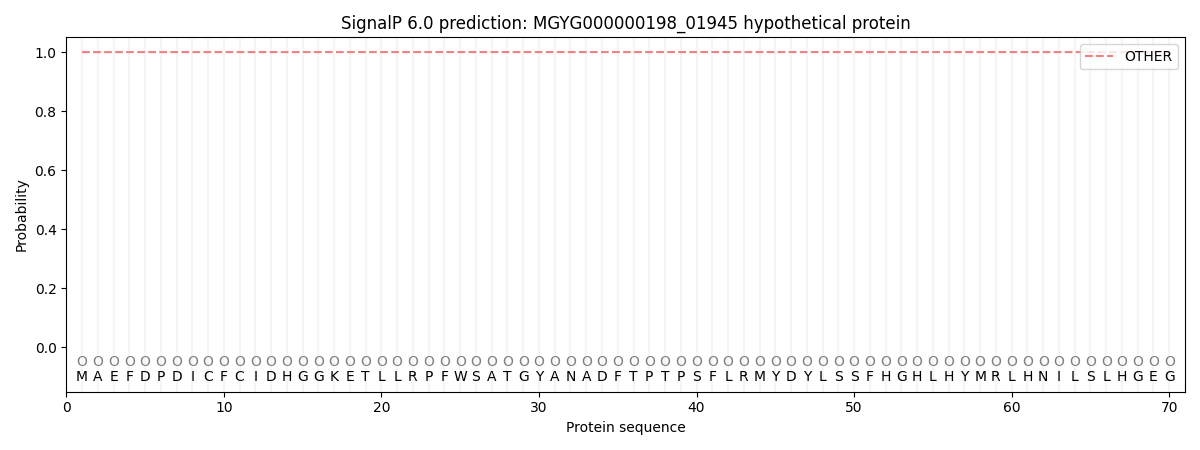
This protein is predicted as OTHER
| Other | SP_Sec_SPI | LIPO_Sec_SPII | TAT_Tat_SPI | TATLIP_Sec_SPII | PILIN_Sec_SPIII |
|---|---|---|---|---|---|
| 1.000036 | 0.000000 | 0.000000 | 0.000000 | 0.000000 | 0.000000 |