You are browsing environment: HUMAN GUT
CAZyme Information: MGYG000000210_01681
You are here: Home > Sequence: MGYG000000210_01681
Basic Information |
Genomic context |
Full Sequence |
Enzyme annotations |
CAZy signature domains |
CDD domains |
CAZyme hits |
PDB hits |
Swiss-Prot hits |
SignalP and Lipop annotations |
TMHMM annotations
Basic Information help
| Species | ||||||||||||
|---|---|---|---|---|---|---|---|---|---|---|---|---|
| Lineage | Bacteria; Actinobacteriota; Coriobacteriia; Coriobacteriales; Coriobacteriaceae; Collinsella; | |||||||||||
| CAZyme ID | MGYG000000210_01681 | |||||||||||
| CAZy Family | GT0 | |||||||||||
| CAZyme Description | UDP-N-acetylglucosamine 2-epimerase | |||||||||||
| CAZyme Property |
|
|||||||||||
| Genome Property |
|
|||||||||||
| Gene Location | Start: 18167; End: 18901 Strand: - | |||||||||||
CDD Domains download full data without filtering help
| Cdd ID | Domain | E-Value | qStart | qEnd | sStart | sEnd | Domain Description |
|---|---|---|---|---|---|---|---|
| cd03786 | GTB_UDP-GlcNAc_2-Epimerase | 4.65e-67 | 2 | 229 | 141 | 365 | UDP-N-acetylglucosamine 2-epimerase and similar proteins. Bacterial members of the UDP-N-Acetylglucosamine (GlcNAc) 2-Epimerase family (EC 5.1.3.14) are known to catalyze the reversible interconversion of UDP-GlcNAc and UDP-N-acetylmannosamine (UDP-ManNAc). The enzyme serves to produce an activated form of ManNAc residues (UDP-ManNAc) for use in the biosynthesis of a variety of cell surface polysaccharides; The mammalian enzyme is bifunctional, catalyzing both the inversion of stereochemistry at C-2 and the hydrolysis of the UDP-sugar linkage to generate free ManNAc. It also catalyzes the phosphorylation of ManNAc to generate ManNAc 6-phosphate, a precursor to salic acids. In mammals, sialic acids are found at the termini of oligosaccharides in a large variety of cell surface glycoconjugates and are key mediators of cell-cell recognition events. Mutations in human members of this family have been associated with Sialuria, a rare disease caused by the disorders of sialic acid metabolism. This family belongs to the GT-B structural superfamily of glycoslytransferases, which have characteristic N- and C-terminal domains each containing a typical Rossmann fold. The two domains have high structural homology despite minimal sequence homology. The large cleft that separates the two domains includes the catalytic center and permits a high degree of flexibility. |
| COG0381 | WecB | 2.33e-64 | 2 | 243 | 145 | 383 | UDP-N-acetylglucosamine 2-epimerase [Cell wall/membrane/envelope biogenesis]. |
CAZyme Hits help
| Hit ID | E-Value | Query Start | Query End | Hit Start | Hit End |
|---|---|---|---|---|---|
| AOP03843.1 | 3.92e-108 | 1 | 242 | 162 | 402 |
| AOP02687.1 | 3.92e-108 | 1 | 242 | 162 | 402 |
| AOP03732.1 | 3.92e-108 | 1 | 242 | 162 | 402 |
| AOP02662.1 | 3.92e-108 | 1 | 242 | 162 | 402 |
| AOP03614.1 | 3.92e-108 | 1 | 242 | 162 | 402 |
PDB Hits download full data without filtering help
| Hit ID | E-Value | Query Start | Query End | Hit Start | Hit End | Description |
|---|---|---|---|---|---|---|
| 4HWG_A | 8.85e-52 | 2 | 243 | 146 | 385 | Structureof UDP-N-acetylglucosamine 2-epimerase from Rickettsia bellii [Rickettsia bellii RML369-C] |
| 4NEQ_A | 4.19e-19 | 2 | 195 | 139 | 334 | Thestructure of UDP-GlcNAc 2-epimerase from Methanocaldococcus jannaschii [Methanocaldococcus jannaschii DSM 2661],4NES_A Crystal structure of Methanocaldococcus jannaschii UDP-GlcNAc 2-epimerase in complex with UDP-GlcNAc and UDP [Methanocaldococcus jannaschii DSM 2661] |
| 1F6D_A | 4.00e-09 | 10 | 230 | 149 | 371 | TheStructure Of Udp-N-Acetylglucosamine 2-Epimerase From E. Coli. [Escherichia coli],1F6D_B The Structure Of Udp-N-Acetylglucosamine 2-Epimerase From E. Coli. [Escherichia coli],1F6D_C The Structure Of Udp-N-Acetylglucosamine 2-Epimerase From E. Coli. [Escherichia coli],1F6D_D The Structure Of Udp-N-Acetylglucosamine 2-Epimerase From E. Coli. [Escherichia coli] |
| 1VGV_A | 4.06e-09 | 10 | 230 | 149 | 371 | Crystalstructure of UDP-N-acetylglucosamine_2 epimerase [Escherichia coli],1VGV_B Crystal structure of UDP-N-acetylglucosamine_2 epimerase [Escherichia coli],1VGV_C Crystal structure of UDP-N-acetylglucosamine_2 epimerase [Escherichia coli],1VGV_D Crystal structure of UDP-N-acetylglucosamine_2 epimerase [Escherichia coli] |
| 3DZC_A | 1.08e-07 | 2 | 228 | 166 | 394 | 2.35Angstrom resolution structure of WecB (VC0917), a UDP-N-acetylglucosamine 2-epimerase from Vibrio cholerae. [Vibrio cholerae],3DZC_B 2.35 Angstrom resolution structure of WecB (VC0917), a UDP-N-acetylglucosamine 2-epimerase from Vibrio cholerae. [Vibrio cholerae] |
Swiss-Prot Hits download full data without filtering help
| Hit ID | E-Value | Query Start | Query End | Hit Start | Hit End | Description |
|---|---|---|---|---|---|---|
| Q6LZC4 | 1.23e-19 | 2 | 208 | 138 | 346 | UDP-N-acetylglucosamine 2-epimerase OS=Methanococcus maripaludis (strain S2 / LL) OX=267377 GN=wecB PE=1 SV=1 |
| Q58899 | 2.15e-18 | 2 | 195 | 139 | 334 | UDP-N-acetylglucosamine 2-epimerase OS=Methanocaldococcus jannaschii (strain ATCC 43067 / DSM 2661 / JAL-1 / JCM 10045 / NBRC 100440) OX=243232 GN=wecB PE=1 SV=1 |
| Q9X0C4 | 1.07e-12 | 29 | 236 | 168 | 372 | Putative UDP-N-acetylglucosamine 2-epimerase OS=Thermotoga maritima (strain ATCC 43589 / DSM 3109 / JCM 10099 / NBRC 100826 / MSB8) OX=243274 GN=TM_1034 PE=3 SV=1 |
| P58600 | 6.55e-12 | 2 | 231 | 142 | 373 | Probable UDP-N-acetylglucosamine 2-epimerase OS=Ralstonia solanacearum (strain GMI1000) OX=267608 GN=epsC PE=3 SV=1 |
| P52641 | 8.87e-12 | 2 | 231 | 142 | 373 | Probable UDP-N-acetylglucosamine 2-epimerase OS=Ralstonia solanacearum OX=305 GN=epsC PE=3 SV=2 |
SignalP and Lipop Annotations help
This protein is predicted as OTHER
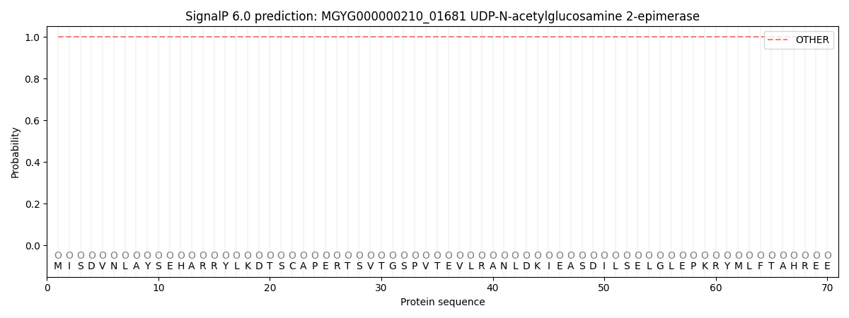
| Other | SP_Sec_SPI | LIPO_Sec_SPII | TAT_Tat_SPI | TATLIP_Sec_SPII | PILIN_Sec_SPIII |
|---|---|---|---|---|---|
| 1.000055 | 0.000000 | 0.000000 | 0.000000 | 0.000000 | 0.000000 |