You are browsing environment: HUMAN GUT
CAZyme Information: MGYG000000223_01797
You are here: Home > Sequence: MGYG000000223_01797
Basic Information |
Genomic context |
Full Sequence |
Enzyme annotations |
CAZy signature domains |
CDD domains |
CAZyme hits |
PDB hits |
Swiss-Prot hits |
SignalP and Lipop annotations |
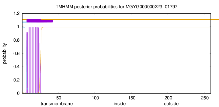
TMHMM annotations
Basic Information help
| Species | NSJ-61 sp003433845 | |||||||||||
|---|---|---|---|---|---|---|---|---|---|---|---|---|
| Lineage | Bacteria; Firmicutes; Bacilli; Erysipelotrichales; Erysipelotrichaceae; NSJ-61; NSJ-61 sp003433845 | |||||||||||
| CAZyme ID | MGYG000000223_01797 | |||||||||||
| CAZy Family | CE4 | |||||||||||
| CAZyme Description | hypothetical protein | |||||||||||
| CAZyme Property |
|
|||||||||||
| Genome Property |
|
|||||||||||
| Gene Location | Start: 51698; End: 52477 Strand: + | |||||||||||
CAZyme Signature Domains help
| Family | Start | End | Evalue | family coverage |
|---|---|---|---|---|
| CE4 | 91 | 220 | 1.4e-19 | 0.9153846153846154 |
CDD Domains download full data without filtering help
| Cdd ID | Domain | E-Value | qStart | qEnd | sStart | sEnd | Domain Description |
|---|---|---|---|---|---|---|---|
| cd10966 | CE4_yadE_5s | 2.19e-45 | 93 | 253 | 1 | 164 | Putative catalytic polysaccharide deacetylase domain of uncharacterized protein yadE and similar proteins. This family contains an uncharacterized protein yadE from Escherichia coli and its bacterial homologs. Although its molecular function remains unknown, yadE shows high sequence similarity with the catalytic NodB homology domain of outer membrane lipoprotein PgaB and the surface-attached protein intercellular adhesion protein IcaB. Both PgaB and IcaB are essential in bacterial biofilm formation. |
| cd10969 | CE4_Ecf1_like_5s | 8.21e-29 | 61 | 248 | 3 | 206 | Putative catalytic NodB homology domain of a hypothetical protein Ecf1 from Escherichia coli and similar proteins. This family contains a hypothetical protein Ecf1 from Escherichia coli and its prokaryotic homologs. Although their biochemical properties remain to be determined, members in this family contain a conserved domain with a 5-stranded beta/alpha barrel, which is similar to the catalytic NodB homology domain of rhizobial NodB-like proteins, belonging to the larger carbohydrate esterase 4 (CE4) superfamily. |
| cd10918 | CE4_NodB_like_5s_6s | 2.38e-26 | 96 | 246 | 1 | 156 | Putative catalytic NodB homology domain of PgaB, IcaB, and similar proteins which consist of a deformed (beta/alpha)8 barrel fold with 5- or 6-strands. This family belongs to the large and functionally diverse carbohydrate esterase 4 (CE4) superfamily, whose members show strong sequence similarity with some variability due to their distinct carbohydrate substrates. It includes bacterial poly-beta-1,6-N-acetyl-D-glucosamine N-deacetylase PgaB, hemin storage system HmsF protein in gram-negative species, intercellular adhesion proteins IcaB, and many uncharacterized prokaryotic polysaccharide deacetylases. It also includes a putative polysaccharide deacetylase YxkH encoded by the Bacillus subtilis yxkH gene, which is one of six polysaccharide deacetylase gene homologs present in the Bacillus subtilis genome. Sequence comparison shows all family members contain a conserved domain similar to the catalytic NodB homology domain of rhizobial NodB-like proteins, which consists of a deformed (beta/alpha)8 barrel fold with 6 or 7 strands. However, in this family, most proteins have 5 strands and some have 6 strands. Moreover, long insertions are found in many family members, whose function remains unknown. |
| cd10973 | CE4_DAC_u4_5s | 2.03e-16 | 95 | 245 | 1 | 154 | Putative catalytic NodB homology domain of uncharacterized bacterial polysaccharide deacetylases which consist of a 5-stranded beta/alpha barrel. This family contains many uncharacterized bacterial polysaccharide deacetylases. Although their biological functions remain unknown, all members of the family are predicted to contain a conserved domain with a 5-stranded beta/alpha barrel, which is similar to the catalytic NodB homology domain of rhizobial NodB-like proteins, belonging to the larger carbohydrate esterase 4 (CE4) superfamily. |
| cd10965 | CE4_IcaB_5s | 3.77e-16 | 93 | 248 | 1 | 163 | Putative catalytic polysaccharide deacetylase domain of bacterial intercellular adhesion protein IcaB and similar proteins. The family is represented by the surface-attached protein intercellular adhesion protein IcaB (Poly-beta-1,6-N-acetyl-D-glucosamine N-deacetylase, EC 3.5.1.-), encoded by Staphylococcus epidermidis icaB gene from the icaABC gene cluster that is involved in the synthesis of polysaccharide intercellular adhesin (PIA), which is located mainly on the cell surface. IcaB is a secreted, cell wall-associated protein that plays a crucial role in exopolysaccharide modification in bacterial biofilm formation. It catalyzes the N-deacetylation of poly-beta-1,6-N-acetyl-D-glucosamine (PNAG, also referred to as PIA), a biofilm adhesin polysaccharide. IcaB shows high homology to the N-terminal NodB homology domain of Escherichia coli PgaB. At this point, they are classified in the same family. |
CAZyme Hits help
| Hit ID | E-Value | Query Start | Query End | Hit Start | Hit End |
|---|---|---|---|---|---|
| AEE91926.1 | 9.20e-27 | 27 | 259 | 44 | 279 |
| CCP26752.1 | 9.20e-27 | 27 | 259 | 44 | 279 |
| ADU29695.1 | 1.96e-25 | 28 | 257 | 70 | 308 |
| AFS79569.1 | 3.55e-21 | 10 | 250 | 3 | 264 |
| ANS75271.1 | 2.78e-20 | 32 | 257 | 36 | 253 |
PDB Hits download full data without filtering help
| Hit ID | E-Value | Query Start | Query End | Hit Start | Hit End | Description |
|---|---|---|---|---|---|---|
| 4WCJ_A | 7.48e-13 | 23 | 222 | 28 | 228 | Structureof IcaB from Ammonifex degensii [Ammonifex degensii KC4] |
| 3VUS_A | 4.82e-08 | 39 | 257 | 14 | 264 | Escherichiacoli PgaB N-terminal domain [Escherichia coli K-12],3VUS_B Escherichia coli PgaB N-terminal domain [Escherichia coli K-12] |
| 4U10_A | 5.15e-08 | 31 | 226 | 3 | 234 | Probingthe structure and mechanism of de-N-acetylase from aggregatibacter actinomycetemcomitans [Aggregatibacter actinomycetemcomitans],4U10_B Probing the structure and mechanism of de-N-acetylase from aggregatibacter actinomycetemcomitans [Aggregatibacter actinomycetemcomitans] |
| 4F9D_A | 8.85e-08 | 39 | 257 | 18 | 268 | Structureof Escherichia coli PgaB 42-655 in complex with nickel [Escherichia coli K-12],4F9D_B Structure of Escherichia coli PgaB 42-655 in complex with nickel [Escherichia coli K-12] |
| 4F9J_A | 2.13e-07 | 39 | 257 | 18 | 268 | Structureof Escherichia coli PgaB 42-655 in complex with iron [Escherichia coli K-12],4F9J_B Structure of Escherichia coli PgaB 42-655 in complex with iron [Escherichia coli K-12] |
Swiss-Prot Hits download full data without filtering help
| Hit ID | E-Value | Query Start | Query End | Hit Start | Hit End | Description |
|---|---|---|---|---|---|---|
| P31666 | 3.66e-24 | 17 | 257 | 156 | 403 | Uncharacterized protein YadE OS=Escherichia coli (strain K12) OX=83333 GN=yadE PE=3 SV=2 |
| P94361 | 2.79e-17 | 27 | 258 | 60 | 276 | Putative polysaccharide deacetylase YxkH OS=Bacillus subtilis (strain 168) OX=224308 GN=yxkH PE=3 SV=1 |
| P75906 | 4.97e-07 | 39 | 257 | 55 | 305 | Poly-beta-1,6-N-acetyl-D-glucosamine N-deacetylase OS=Escherichia coli (strain K12) OX=83333 GN=pgaB PE=1 SV=1 |
| Q8XAR3 | 6.66e-07 | 39 | 257 | 55 | 305 | Poly-beta-1,6-N-acetyl-D-glucosamine N-deacetylase OS=Escherichia coli O157:H7 OX=83334 GN=pgaB PE=3 SV=1 |
SignalP and Lipop Annotations help
This protein is predicted as OTHER

| Other | SP_Sec_SPI | LIPO_Sec_SPII | TAT_Tat_SPI | TATLIP_Sec_SPII | PILIN_Sec_SPIII |
|---|---|---|---|---|---|
| 0.971631 | 0.021992 | 0.006004 | 0.000075 | 0.000054 | 0.000273 |