You are browsing environment: HUMAN GUT
CAZyme Information: MGYG000000247_00629
You are here: Home > Sequence: MGYG000000247_00629
Basic Information |
Genomic context |
Full Sequence |
Enzyme annotations |
CAZy signature domains |
CDD domains |
CAZyme hits |
PDB hits |
Swiss-Prot hits |
SignalP and Lipop annotations |
TMHMM annotations
Basic Information help
| Species | Collinsella intestinalis | |||||||||||
|---|---|---|---|---|---|---|---|---|---|---|---|---|
| Lineage | Bacteria; Actinobacteriota; Coriobacteriia; Coriobacteriales; Coriobacteriaceae; Collinsella; Collinsella intestinalis | |||||||||||
| CAZyme ID | MGYG000000247_00629 | |||||||||||
| CAZy Family | GT4 | |||||||||||
| CAZyme Description | GDP-mannose-dependent monoacylated alpha-(1-6)-phosphatidylinositol monomannoside mannosyltransferase | |||||||||||
| CAZyme Property |
|
|||||||||||
| Genome Property |
|
|||||||||||
| Gene Location | Start: 730307; End: 731437 Strand: + | |||||||||||
CDD Domains download full data without filtering help
| Cdd ID | Domain | E-Value | qStart | qEnd | sStart | sEnd | Domain Description |
|---|---|---|---|---|---|---|---|
| cd03801 | GT4_PimA-like | 1.11e-55 | 4 | 375 | 2 | 365 | phosphatidyl-myo-inositol mannosyltransferase. This family is most closely related to the GT4 family of glycosyltransferases and named after PimA in Propionibacterium freudenreichii, which is involved in the biosynthesis of phosphatidyl-myo-inositol mannosides (PIM) which are early precursors in the biosynthesis of lipomannans (LM) and lipoarabinomannans (LAM), and catalyzes the addition of a mannosyl residue from GDP-D-mannose (GDP-Man) to the position 2 of the carrier lipid phosphatidyl-myo-inositol (PI) to generate a phosphatidyl-myo-inositol bearing an alpha-1,2-linked mannose residue (PIM1). Glycosyltransferases catalyze the transfer of sugar moieties from activated donor molecules to specific acceptor molecules, forming glycosidic bonds. The acceptor molecule can be a lipid, a protein, a heterocyclic compound, or another carbohydrate residue. This group of glycosyltransferases is most closely related to the previously defined glycosyltransferase family 1 (GT1). The members of this family may transfer UDP, ADP, GDP, or CMP linked sugars. The diverse enzymatic activities among members of this family reflect a wide range of biological functions. The protein structure available for this family has the GTB topology, one of the two protein topologies observed for nucleotide-sugar-dependent glycosyltransferases. GTB proteins have distinct N- and C- terminal domains each containing a typical Rossmann fold. The two domains have high structural homology despite minimal sequence homology. The large cleft that separates the two domains includes the catalytic center and permits a high degree of flexibility. The members of this family are found mainly in certain bacteria and archaea. |
| COG0438 | RfaB | 7.62e-45 | 4 | 376 | 3 | 375 | Glycosyltransferase involved in cell wall bisynthesis [Cell wall/membrane/envelope biogenesis]. |
| cd03814 | GT4-like | 4.05e-39 | 4 | 376 | 2 | 365 | glycosyltransferase family 4 proteins. This family is most closely related to the GT4 family of glycosyltransferases and includes a sequence annotated as alpha-D-mannose-alpha(1-6)phosphatidyl myo-inositol monomannoside transferase from Bacillus halodurans. Glycosyltransferases catalyze the transfer of sugar moieties from activated donor molecules to specific acceptor molecules, forming glycosidic bonds. The acceptor molecule can be a lipid, a protein, a heterocyclic compound, or another carbohydrate residue. This group of glycosyltransferases is most closely related to the previously defined glycosyltransferase family 1 (GT1). The members of this family may transfer UDP, ADP, GDP, or CMP linked sugars. The diverse enzymatic activities among members of this family reflect a wide range of biological functions. The protein structure available for this family has the GTB topology, one of the two protein topologies observed for nucleotide-sugar-dependent glycosyltransferases. GTB proteins have distinct N- and C- terminal domains each containing a typical Rossmann fold. The two domains have high structural homology despite minimal sequence homology. The large cleft that separates the two domains includes the catalytic center and permits a high degree of flexibility. The members of this family are found mainly in bacteria and eukaryotes. |
| cd03798 | GT4_WlbH-like | 4.34e-39 | 4 | 376 | 1 | 374 | Bordetella parapertussis WlbH and similar proteins. This family is most closely related to the GT4 family of glycosyltransferases. Staphylococcus aureus CapJ may be involved in capsule polysaccharide biosynthesis. WlbH in Bordetella parapertussis has been shown to be required for the biosynthesis of a trisaccharide that, when attached to the B. pertussis lipopolysaccharide (LPS) core (band B), generates band A LPS. |
| cd03811 | GT4_GT28_WabH-like | 2.42e-37 | 4 | 350 | 4 | 335 | family 4 and family 28 glycosyltransferases similar to Klebsiella WabH. This family is most closely related to the GT1 family of glycosyltransferases. WabH in Klebsiella pneumoniae has been shown to transfer a GlcNAc residue from UDP-GlcNAc onto the acceptor GalUA residue in the cellular outer core. |
CAZyme Hits help
| Hit ID | E-Value | Query Start | Query End | Hit Start | Hit End |
|---|---|---|---|---|---|
| CBL03246.1 | 1.34e-111 | 2 | 368 | 14 | 381 |
| QOY61384.1 | 7.25e-108 | 6 | 376 | 9 | 381 |
| AZH69451.1 | 7.78e-105 | 2 | 368 | 3 | 371 |
| BAN76660.1 | 7.89e-97 | 4 | 364 | 5 | 366 |
| AEB07231.1 | 8.63e-95 | 4 | 376 | 3 | 378 |
PDB Hits download full data without filtering help
| Hit ID | E-Value | Query Start | Query End | Hit Start | Hit End | Description |
|---|---|---|---|---|---|---|
| 2JJM_A | 2.51e-11 | 13 | 351 | 24 | 358 | CrystalStructure of a family GT4 glycosyltransferase from Bacillus anthracis ORF BA1558. [Bacillus anthracis str. Ames],2JJM_B Crystal Structure of a family GT4 glycosyltransferase from Bacillus anthracis ORF BA1558. [Bacillus anthracis str. Ames],2JJM_C Crystal Structure of a family GT4 glycosyltransferase from Bacillus anthracis ORF BA1558. [Bacillus anthracis str. Ames],2JJM_D Crystal Structure of a family GT4 glycosyltransferase from Bacillus anthracis ORF BA1558. [Bacillus anthracis str. Ames],2JJM_E Crystal Structure of a family GT4 glycosyltransferase from Bacillus anthracis ORF BA1558. [Bacillus anthracis str. Ames],2JJM_F Crystal Structure of a family GT4 glycosyltransferase from Bacillus anthracis ORF BA1558. [Bacillus anthracis str. Ames],2JJM_G Crystal Structure of a family GT4 glycosyltransferase from Bacillus anthracis ORF BA1558. [Bacillus anthracis str. Ames],2JJM_H Crystal Structure of a family GT4 glycosyltransferase from Bacillus anthracis ORF BA1558. [Bacillus anthracis str. Ames],2JJM_I Crystal Structure of a family GT4 glycosyltransferase from Bacillus anthracis ORF BA1558. [Bacillus anthracis str. Ames],2JJM_J Crystal Structure of a family GT4 glycosyltransferase from Bacillus anthracis ORF BA1558. [Bacillus anthracis str. Ames],2JJM_K Crystal Structure of a family GT4 glycosyltransferase from Bacillus anthracis ORF BA1558. [Bacillus anthracis str. Ames],2JJM_L Crystal Structure of a family GT4 glycosyltransferase from Bacillus anthracis ORF BA1558. [Bacillus anthracis str. Ames] |
| 3MBO_A | 2.68e-11 | 13 | 351 | 44 | 378 | CrystalStructure of the Glycosyltransferase BaBshA bound with UDP and L-malate [Bacillus anthracis],3MBO_B Crystal Structure of the Glycosyltransferase BaBshA bound with UDP and L-malate [Bacillus anthracis],3MBO_C Crystal Structure of the Glycosyltransferase BaBshA bound with UDP and L-malate [Bacillus anthracis],3MBO_D Crystal Structure of the Glycosyltransferase BaBshA bound with UDP and L-malate [Bacillus anthracis],3MBO_E Crystal Structure of the Glycosyltransferase BaBshA bound with UDP and L-malate [Bacillus anthracis],3MBO_F Crystal Structure of the Glycosyltransferase BaBshA bound with UDP and L-malate [Bacillus anthracis],3MBO_G Crystal Structure of the Glycosyltransferase BaBshA bound with UDP and L-malate [Bacillus anthracis],3MBO_H Crystal Structure of the Glycosyltransferase BaBshA bound with UDP and L-malate [Bacillus anthracis] |
| 6KIH_A | 1.64e-09 | 199 | 368 | 235 | 414 | Sucrose-phosphatesynthase (tll1590) from Thermosynechococcus elongatus [Thermosynechococcus vestitus],6KIH_B Sucrose-phosphate synthase (tll1590) from Thermosynechococcus elongatus [Thermosynechococcus vestitus],6KIH_C Sucrose-phosphate synthase (tll1590) from Thermosynechococcus elongatus [Thermosynechococcus vestitus],6KIH_D Sucrose-phosphate synthase (tll1590) from Thermosynechococcus elongatus [Thermosynechococcus vestitus],6KIH_E Sucrose-phosphate synthase (tll1590) from Thermosynechococcus elongatus [Thermosynechococcus vestitus],6KIH_F Sucrose-phosphate synthase (tll1590) from Thermosynechococcus elongatus [Thermosynechococcus vestitus],6KIH_G Sucrose-phosphate synthase (tll1590) from Thermosynechococcus elongatus [Thermosynechococcus vestitus],6KIH_H Sucrose-phosphate synthase (tll1590) from Thermosynechococcus elongatus [Thermosynechococcus vestitus],6KIH_I Sucrose-phosphate synthase (tll1590) from Thermosynechococcus elongatus [Thermosynechococcus vestitus],6KIH_J Sucrose-phosphate synthase (tll1590) from Thermosynechococcus elongatus [Thermosynechococcus vestitus],6KIH_K Sucrose-phosphate synthase (tll1590) from Thermosynechococcus elongatus [Thermosynechococcus vestitus],6KIH_L Sucrose-phosphate synthase (tll1590) from Thermosynechococcus elongatus [Thermosynechococcus vestitus] |
| 3C4Q_A | 1.14e-08 | 192 | 376 | 208 | 403 | Structureof the retaining glycosyltransferase MshA : The first step in mycothiol biosynthesis. Organism : Corynebacterium glutamicum- Complex with UDP [Corynebacterium glutamicum],3C4Q_B Structure of the retaining glycosyltransferase MshA : The first step in mycothiol biosynthesis. Organism : Corynebacterium glutamicum- Complex with UDP [Corynebacterium glutamicum],3C4V_A Structure of the retaining glycosyltransferase MshA:The first step in mycothiol biosynthesis. Organism: Corynebacterium glutamicum : Complex with UDP and 1L-INS-1-P. [Corynebacterium glutamicum],3C4V_B Structure of the retaining glycosyltransferase MshA:The first step in mycothiol biosynthesis. Organism: Corynebacterium glutamicum : Complex with UDP and 1L-INS-1-P. [Corynebacterium glutamicum] |
| 3C48_A | 1.17e-08 | 192 | 376 | 228 | 423 | Structureof the retaining glycosyltransferase MshA: The first step in mycothiol biosynthesis. Organism: Corynebacterium glutamicum- APO (OPEN) structure. [Corynebacterium glutamicum],3C48_B Structure of the retaining glycosyltransferase MshA: The first step in mycothiol biosynthesis. Organism: Corynebacterium glutamicum- APO (OPEN) structure. [Corynebacterium glutamicum] |
Swiss-Prot Hits download full data without filtering help
| Hit ID | E-Value | Query Start | Query End | Hit Start | Hit End | Description |
|---|---|---|---|---|---|---|
| P37287 | 2.49e-21 | 4 | 317 | 35 | 341 | Phosphatidylinositol N-acetylglucosaminyltransferase subunit A OS=Homo sapiens OX=9606 GN=PIGA PE=1 SV=1 |
| Q64323 | 7.46e-19 | 4 | 317 | 35 | 342 | Phosphatidylinositol N-acetylglucosaminyltransferase subunit A OS=Mus musculus OX=10090 GN=Piga PE=2 SV=1 |
| A1R8N8 | 2.22e-18 | 16 | 375 | 20 | 396 | D-inositol 3-phosphate glycosyltransferase OS=Paenarthrobacter aurescens (strain TC1) OX=290340 GN=mshA PE=3 SV=1 |
| P87172 | 7.16e-18 | 8 | 368 | 3 | 355 | Phosphatidylinositol N-acetylglucosaminyltransferase gpi3 subunit OS=Schizosaccharomyces pombe (strain 972 / ATCC 24843) OX=284812 GN=gpi3 PE=3 SV=1 |
| B8HCF8 | 9.44e-16 | 16 | 375 | 26 | 402 | D-inositol 3-phosphate glycosyltransferase OS=Pseudarthrobacter chlorophenolicus (strain ATCC 700700 / DSM 12829 / CIP 107037 / JCM 12360 / KCTC 9906 / NCIMB 13794 / A6) OX=452863 GN=mshA PE=3 SV=1 |
SignalP and Lipop Annotations help
This protein is predicted as OTHER
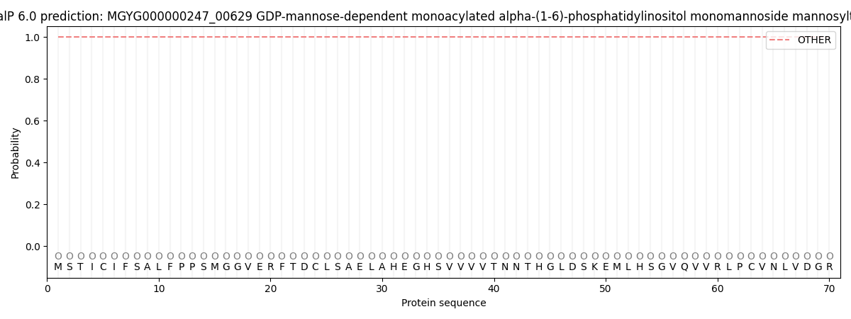
| Other | SP_Sec_SPI | LIPO_Sec_SPII | TAT_Tat_SPI | TATLIP_Sec_SPII | PILIN_Sec_SPIII |
|---|---|---|---|---|---|
| 1.000056 | 0.000001 | 0.000000 | 0.000000 | 0.000000 | 0.000000 |