You are browsing environment: HUMAN GUT
CAZyme Information: MGYG000000247_00633
You are here: Home > Sequence: MGYG000000247_00633
Basic Information |
Genomic context |
Full Sequence |
Enzyme annotations |
CAZy signature domains |
CDD domains |
CAZyme hits |
PDB hits |
Swiss-Prot hits |
SignalP and Lipop annotations |
TMHMM annotations
Basic Information help
| Species | Collinsella intestinalis | |||||||||||
|---|---|---|---|---|---|---|---|---|---|---|---|---|
| Lineage | Bacteria; Actinobacteriota; Coriobacteriia; Coriobacteriales; Coriobacteriaceae; Collinsella; Collinsella intestinalis | |||||||||||
| CAZyme ID | MGYG000000247_00633 | |||||||||||
| CAZy Family | GT2 | |||||||||||
| CAZyme Description | hypothetical protein | |||||||||||
| CAZyme Property |
|
|||||||||||
| Genome Property |
|
|||||||||||
| Gene Location | Start: 736789; End: 739143 Strand: + | |||||||||||
CAZyme Signature Domains help
| Family | Start | End | Evalue | family coverage |
|---|---|---|---|---|
| GT2 | 506 | 688 | 2.1e-21 | 0.9705882352941176 |
| GT2 | 253 | 364 | 2.8e-19 | 0.6588235294117647 |
CDD Domains download full data without filtering help
| Cdd ID | Domain | E-Value | qStart | qEnd | sStart | sEnd | Domain Description |
|---|---|---|---|---|---|---|---|
| cd04184 | GT2_RfbC_Mx_like | 2.18e-83 | 250 | 448 | 1 | 202 | Myxococcus xanthus RfbC like proteins are required for O-antigen biosynthesis. The rfbC gene encodes a predicted protein of 1,276 amino acids, which is required for O-antigen biosynthesis in Myxococcus xanthus. It is a subfamily of Glycosyltransferase Family GT2, which includes diverse families of glycosyl transferases with a common GT-A type structural fold, which has two tightly associated beta/alpha/beta domains that tend to form a continuous central sheet of at least eight beta-strands. These are enzymes that catalyze the transfer of sugar moieties from activated donor molecules to specific acceptor molecules, forming glycosidic bonds. |
| cd04186 | GT_2_like_c | 4.38e-41 | 507 | 730 | 1 | 166 | Subfamily of Glycosyltransferase Family GT2 of unknown function. GT-2 includes diverse families of glycosyltransferases with a common GT-A type structural fold, which has two tightly associated beta/alpha/beta domains that tend to form a continuous central sheet of at least eight beta-strands. These are enzymes that catalyze the transfer of sugar moieties from activated donor molecules to specific acceptor molecules, forming glycosidic bonds. Glycosyltransferases have been classified into more than 90 distinct sequence based families. |
| COG1216 | GT2 | 2.34e-29 | 503 | 736 | 3 | 227 | Glycosyltransferase, GT2 family [Carbohydrate transport and metabolism]. |
| cd06433 | GT_2_WfgS_like | 2.43e-20 | 253 | 453 | 1 | 200 | WfgS and WfeV are involved in O-antigen biosynthesis. Escherichia coli WfgS and Shigella dysenteriae WfeV are glycosyltransferase 2 family enzymes involved in O-antigen biosynthesis. GT-2 enzymes have GT-A type structural fold, which has two tightly associated beta/alpha/beta domains that tend to form a continuous central sheet of at least eight beta-strands. These are enzymes that catalyze the transfer of sugar moieties from activated donor molecules to specific acceptor molecules, forming glycosidic bonds. Glycosyltransferases have been classified into more than 90 distinct sequence based families. |
| pfam00535 | Glycos_transf_2 | 4.14e-19 | 253 | 417 | 1 | 164 | Glycosyl transferase family 2. Diverse family, transferring sugar from UDP-glucose, UDP-N-acetyl- galactosamine, GDP-mannose or CDP-abequose, to a range of substrates including cellulose, dolichol phosphate and teichoic acids. |
CAZyme Hits help
| Hit ID | E-Value | Query Start | Query End | Hit Start | Hit End |
|---|---|---|---|---|---|
| AEB07226.1 | 2.99e-289 | 2 | 768 | 23 | 789 |
| ACV56006.1 | 5.74e-191 | 16 | 770 | 60 | 818 |
| AZH69456.1 | 3.05e-185 | 20 | 775 | 57 | 814 |
| QOY61387.1 | 1.11e-181 | 38 | 775 | 61 | 800 |
| ATP53720.1 | 1.69e-181 | 20 | 775 | 57 | 814 |
PDB Hits download full data without filtering help
| Hit ID | E-Value | Query Start | Query End | Hit Start | Hit End | Description |
|---|---|---|---|---|---|---|
| 5HEA_A | 2.81e-09 | 249 | 396 | 4 | 145 | CgTstructure in hexamer [Streptococcus parasanguinis FW213],5HEA_B CgT structure in hexamer [Streptococcus parasanguinis FW213],5HEA_C CgT structure in hexamer [Streptococcus parasanguinis FW213],5HEC_A CgT structure in dimer [Streptococcus parasanguinis FW213],5HEC_B CgT structure in dimer [Streptococcus parasanguinis FW213] |
| 3L7I_A | 7.60e-08 | 251 | 447 | 3 | 205 | Structureof the Wall Teichoic Acid Polymerase TagF [Staphylococcus epidermidis RP62A],3L7I_B Structure of the Wall Teichoic Acid Polymerase TagF [Staphylococcus epidermidis RP62A],3L7I_C Structure of the Wall Teichoic Acid Polymerase TagF [Staphylococcus epidermidis RP62A],3L7I_D Structure of the Wall Teichoic Acid Polymerase TagF [Staphylococcus epidermidis RP62A] |
| 3L7J_A | 7.60e-08 | 251 | 447 | 3 | 205 | ChainA, Teichoic acid biosynthesis protein F [Staphylococcus epidermidis RP62A],3L7J_B Chain B, Teichoic acid biosynthesis protein F [Staphylococcus epidermidis RP62A],3L7J_C Chain C, Teichoic acid biosynthesis protein F [Staphylococcus epidermidis RP62A],3L7J_D Chain D, Teichoic acid biosynthesis protein F [Staphylococcus epidermidis RP62A],3L7K_A Chain A, Teichoic acid biosynthesis protein F [Staphylococcus epidermidis RP62A],3L7K_B Chain B, Teichoic acid biosynthesis protein F [Staphylococcus epidermidis RP62A],3L7K_C Chain C, Teichoic acid biosynthesis protein F [Staphylococcus epidermidis RP62A],3L7K_D Chain D, Teichoic acid biosynthesis protein F [Staphylococcus epidermidis RP62A],3L7L_A Chain A, Teichoic acid biosynthesis protein F [Staphylococcus epidermidis RP62A],3L7L_B Chain B, Teichoic acid biosynthesis protein F [Staphylococcus epidermidis RP62A],3L7L_C Chain C, Teichoic acid biosynthesis protein F [Staphylococcus epidermidis RP62A],3L7L_D Chain D, Teichoic acid biosynthesis protein F [Staphylococcus epidermidis RP62A] |
| 3L7M_A | 7.60e-08 | 251 | 447 | 3 | 205 | ChainA, Teichoic acid biosynthesis protein F [Staphylococcus epidermidis RP62A],3L7M_B Chain B, Teichoic acid biosynthesis protein F [Staphylococcus epidermidis RP62A],3L7M_C Chain C, Teichoic acid biosynthesis protein F [Staphylococcus epidermidis RP62A],3L7M_D Chain D, Teichoic acid biosynthesis protein F [Staphylococcus epidermidis RP62A] |
| 2Z86_A | 8.34e-07 | 250 | 480 | 375 | 603 | Crystalstructure of chondroitin polymerase from Escherichia coli strain K4 (K4CP) complexed with UDP-GlcUA and UDP [Escherichia coli],2Z86_B Crystal structure of chondroitin polymerase from Escherichia coli strain K4 (K4CP) complexed with UDP-GlcUA and UDP [Escherichia coli],2Z86_C Crystal structure of chondroitin polymerase from Escherichia coli strain K4 (K4CP) complexed with UDP-GlcUA and UDP [Escherichia coli],2Z86_D Crystal structure of chondroitin polymerase from Escherichia coli strain K4 (K4CP) complexed with UDP-GlcUA and UDP [Escherichia coli] |
Swiss-Prot Hits download full data without filtering help
| Hit ID | E-Value | Query Start | Query End | Hit Start | Hit End | Description |
|---|---|---|---|---|---|---|
| P55465 | 3.04e-117 | 249 | 770 | 365 | 886 | Uncharacterized protein y4gI OS=Sinorhizobium fredii (strain NBRC 101917 / NGR234) OX=394 GN=NGR_a03550 PE=4 SV=1 |
| Q50864 | 4.35e-93 | 250 | 774 | 319 | 833 | O-antigen biosynthesis protein RfbC OS=Myxococcus xanthus OX=34 GN=rfbC PE=4 SV=1 |
| Q1RIM7 | 7.52e-13 | 239 | 595 | 2 | 379 | Uncharacterized glycosyltransferase RBE_0706 OS=Rickettsia bellii (strain RML369-C) OX=336407 GN=RBE_0706 PE=3 SV=1 |
| P9WMY3 | 1.26e-08 | 518 | 737 | 18 | 244 | N-acetylglucosaminyl-diphospho-decaprenol L-rhamnosyltransferase OS=Mycobacterium tuberculosis (strain ATCC 25618 / H37Rv) OX=83332 GN=wbbL PE=1 SV=2 |
| P9WMY2 | 1.26e-08 | 518 | 737 | 18 | 244 | N-acetylglucosaminyl-diphospho-decaprenol L-rhamnosyltransferase OS=Mycobacterium tuberculosis (strain CDC 1551 / Oshkosh) OX=83331 GN=wbbL PE=3 SV=2 |
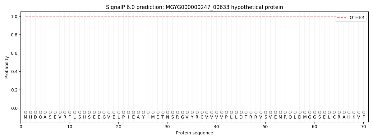
SignalP and Lipop Annotations help
This protein is predicted as OTHER
| Other | SP_Sec_SPI | LIPO_Sec_SPII | TAT_Tat_SPI | TATLIP_Sec_SPII | PILIN_Sec_SPIII |
|---|---|---|---|---|---|
| 1.000073 | 0.000000 | 0.000000 | 0.000000 | 0.000000 | 0.000000 |