You are browsing environment: HUMAN GUT
CAZyme Information: MGYG000000277_01354
You are here: Home > Sequence: MGYG000000277_01354
Basic Information |
Genomic context |
Full Sequence |
Enzyme annotations |
CAZy signature domains |
CDD domains |
CAZyme hits |
PDB hits |
Swiss-Prot hits |
SignalP and Lipop annotations |
TMHMM annotations
Basic Information help
| Species | Streptococcus mutans | |||||||||||
|---|---|---|---|---|---|---|---|---|---|---|---|---|
| Lineage | Bacteria; Firmicutes; Bacilli; Lactobacillales; Streptococcaceae; Streptococcus; Streptococcus mutans | |||||||||||
| CAZyme ID | MGYG000000277_01354 | |||||||||||
| CAZy Family | GH70 | |||||||||||
| CAZyme Description | Glucosyltransferase-SI | |||||||||||
| CAZyme Property |
|
|||||||||||
| Genome Property |
|
|||||||||||
| Gene Location | Start: 8; End: 1495 Strand: + | |||||||||||
CDD Domains download full data without filtering help
| Cdd ID | Domain | E-Value | qStart | qEnd | sStart | sEnd | Domain Description |
|---|---|---|---|---|---|---|---|
| pfam02324 | Glyco_hydro_70 | 6.62e-69 | 1 | 143 | 665 | 804 | Glycosyl hydrolase family 70. Members of this family belong to glycosyl hydrolase family 70 Glucosyltransferases or sucrose 6-glycosyl transferases (GTF-S) catalyze the transfer of D-glucopyramnosyl units from sucrose onto acceptor molecules, EC:2.4.1.5. This family roughly corresponds to the N-terminal catalytic domain of the enzyme. Members of this family also contain the Putative cell wall binding domain pfam01473, which corresponds with the C-terminal glucan-binding domain. |
| TIGR04035 | glucan_65_rpt | 4.13e-18 | 296 | 356 | 3 | 62 | glucan-binding repeat. This model describes a region of about 63 amino acids that is composed of three repeats of a more broadly distributed family of shorter repeats modeled by pfam01473. While the shorter repeats are often associated with choline binding (and therefore with cell wall binding), the longer repeat described here represents a subgroup of repeat sequences associated with glucan binding, as found in a number glycosylhydrolases. Shah, et al. describe a repeat consensus, WYYFDANGKAVTGAQTINGQTLYFDQDGKQVKG, that corresponds to half of the repeat as modeled here and one and a half copies of the repeat as modeled by pfam01473. |
| TIGR04035 | glucan_65_rpt | 2.13e-16 | 361 | 421 | 3 | 62 | glucan-binding repeat. This model describes a region of about 63 amino acids that is composed of three repeats of a more broadly distributed family of shorter repeats modeled by pfam01473. While the shorter repeats are often associated with choline binding (and therefore with cell wall binding), the longer repeat described here represents a subgroup of repeat sequences associated with glucan binding, as found in a number glycosylhydrolases. Shah, et al. describe a repeat consensus, WYYFDANGKAVTGAQTINGQTLYFDQDGKQVKG, that corresponds to half of the repeat as modeled here and one and a half copies of the repeat as modeled by pfam01473. |
| NF033930 | pneumo_PspA | 2.43e-16 | 166 | 450 | 452 | 657 | pneumococcal surface protein A. The pneumococcal surface protein proteins, found in Streptococcus pneumoniae, are repetitive, with patterns of localized high sequence identity across pairs of proteins given different specific names that recombination may be presumed. This protein, PspA, has an N-terminal region that lacks a cross-wall-targeting YSIRK type extended signal peptide, in contrast to the closely related choline-binding protein CbpA which has a similar C-terminus but a YSIRK-containing region at the N-terminus. |
| TIGR04035 | glucan_65_rpt | 1.29e-14 | 167 | 226 | 1 | 60 | glucan-binding repeat. This model describes a region of about 63 amino acids that is composed of three repeats of a more broadly distributed family of shorter repeats modeled by pfam01473. While the shorter repeats are often associated with choline binding (and therefore with cell wall binding), the longer repeat described here represents a subgroup of repeat sequences associated with glucan binding, as found in a number glycosylhydrolases. Shah, et al. describe a repeat consensus, WYYFDANGKAVTGAQTINGQTLYFDQDGKQVKG, that corresponds to half of the repeat as modeled here and one and a half copies of the repeat as modeled by pfam01473. |
CAZyme Hits help
| Hit ID | E-Value | Query Start | Query End | Hit Start | Hit End |
|---|---|---|---|---|---|
| BAA26102.1 | 0.0 | 1 | 495 | 961 | 1455 |
| AYO47739.1 | 0.0 | 1 | 495 | 961 | 1455 |
| AFM81412.1 | 0.0 | 1 | 495 | 961 | 1455 |
| BAH88043.1 | 0.0 | 1 | 495 | 961 | 1455 |
| BAA26120.1 | 0.0 | 1 | 495 | 961 | 1455 |
PDB Hits download full data without filtering help
| Hit ID | E-Value | Query Start | Query End | Hit Start | Hit End | Description |
|---|---|---|---|---|---|---|
| 3AIB_A | 2.69e-76 | 1 | 127 | 718 | 844 | CrystalStructure of Glucansucrase [Streptococcus mutans],3AIB_B Crystal Structure of Glucansucrase [Streptococcus mutans],3AIB_C Crystal Structure of Glucansucrase [Streptococcus mutans],3AIB_D Crystal Structure of Glucansucrase [Streptococcus mutans],3AIB_E Crystal Structure of Glucansucrase [Streptococcus mutans],3AIB_F Crystal Structure of Glucansucrase [Streptococcus mutans],3AIB_G Crystal Structure of Glucansucrase [Streptococcus mutans],3AIB_H Crystal Structure of Glucansucrase [Streptococcus mutans],3AIC_A Crystal Structure of Glucansucrase from Streptococcus mutans [Streptococcus mutans],3AIC_B Crystal Structure of Glucansucrase from Streptococcus mutans [Streptococcus mutans],3AIC_C Crystal Structure of Glucansucrase from Streptococcus mutans [Streptococcus mutans],3AIC_D Crystal Structure of Glucansucrase from Streptococcus mutans [Streptococcus mutans],3AIC_E Crystal Structure of Glucansucrase from Streptococcus mutans [Streptococcus mutans],3AIC_F Crystal Structure of Glucansucrase from Streptococcus mutans [Streptococcus mutans],3AIC_G Crystal Structure of Glucansucrase from Streptococcus mutans [Streptococcus mutans],3AIC_H Crystal Structure of Glucansucrase from Streptococcus mutans [Streptococcus mutans],3AIE_A Crystal Structure of glucansucrase from Streptococcus mutans [Streptococcus mutans],3AIE_B Crystal Structure of glucansucrase from Streptococcus mutans [Streptococcus mutans],3AIE_C Crystal Structure of glucansucrase from Streptococcus mutans [Streptococcus mutans],3AIE_D Crystal Structure of glucansucrase from Streptococcus mutans [Streptococcus mutans],3AIE_E Crystal Structure of glucansucrase from Streptococcus mutans [Streptococcus mutans],3AIE_F Crystal Structure of glucansucrase from Streptococcus mutans [Streptococcus mutans],3AIE_G Crystal Structure of glucansucrase from Streptococcus mutans [Streptococcus mutans],3AIE_H Crystal Structure of glucansucrase from Streptococcus mutans [Streptococcus mutans] |
| 6SYQ_B | 7.68e-54 | 1 | 197 | 937 | 1142 | ChainB, Alternansucrase [Leuconostoc mesenteroides] |
| 6SYQ_A | 8.09e-54 | 1 | 197 | 1026 | 1231 | ChainA, Alternansucrase [Leuconostoc mesenteroides] |
| 6SZI_A | 8.10e-54 | 1 | 197 | 1029 | 1234 | ChainA, Alternansucrase [Leuconostoc mesenteroides],6SZI_B Chain B, Alternansucrase [Leuconostoc mesenteroides],6T16_A Chain A, Alternansucrase [Leuconostoc mesenteroides],6T16_B Chain B, Alternansucrase [Leuconostoc mesenteroides],6T18_A ASR Alternansucrase in complex with oligoalternan [Leuconostoc mesenteroides],6T18_B ASR Alternansucrase in complex with oligoalternan [Leuconostoc mesenteroides],6T1P_A Chain A, Alternansucrase [Leuconostoc mesenteroides],6T1P_B Chain B, Alternansucrase [Leuconostoc mesenteroides] |
| 6HVG_A | 8.55e-54 | 1 | 197 | 1161 | 1366 | ChainA, Alternansucrase [Leuconostoc mesenteroides],6HVG_B Chain B, Alternansucrase [Leuconostoc mesenteroides] |
Swiss-Prot Hits download full data without filtering help
| Hit ID | E-Value | Query Start | Query End | Hit Start | Hit End | Description |
|---|---|---|---|---|---|---|
| P13470 | 0.0 | 1 | 495 | 961 | 1455 | Glucosyltransferase-SI OS=Streptococcus mutans serotype c (strain ATCC 700610 / UA159) OX=210007 GN=gtfC PE=1 SV=2 |
| P08987 | 4.98e-170 | 1 | 451 | 935 | 1383 | Glucosyltransferase-I OS=Streptococcus mutans serotype c (strain ATCC 700610 / UA159) OX=210007 GN=gtfB PE=3 SV=3 |
| P11001 | 2.79e-144 | 1 | 432 | 938 | 1365 | Glucosyltransferase-I OS=Streptococcus downei OX=1317 GN=gtfI PE=3 SV=1 |
| P27470 | 9.80e-141 | 1 | 454 | 932 | 1381 | Glucosyltransferase-I OS=Streptococcus downei OX=1317 PE=3 SV=1 |
| P49331 | 1.34e-132 | 1 | 450 | 966 | 1412 | Glucosyltransferase-S OS=Streptococcus mutans serotype c (strain ATCC 700610 / UA159) OX=210007 GN=gtfD PE=3 SV=3 |
SignalP and Lipop Annotations help
This protein is predicted as OTHER
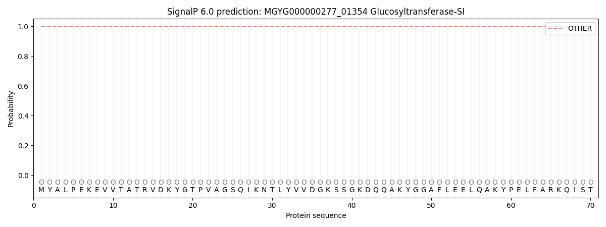
| Other | SP_Sec_SPI | LIPO_Sec_SPII | TAT_Tat_SPI | TATLIP_Sec_SPII | PILIN_Sec_SPIII |
|---|---|---|---|---|---|
| 1.000053 | 0.000000 | 0.000000 | 0.000000 | 0.000000 | 0.000000 |