You are browsing environment: HUMAN GUT
CAZyme Information: MGYG000000298_00293
You are here: Home > Sequence: MGYG000000298_00293
Basic Information |
Genomic context |
Full Sequence |
Enzyme annotations |
CAZy signature domains |
CDD domains |
CAZyme hits |
PDB hits |
Swiss-Prot hits |
SignalP and Lipop annotations |
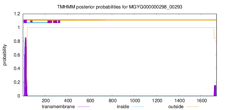
TMHMM annotations
Basic Information help
| Species | ||||||||||||
|---|---|---|---|---|---|---|---|---|---|---|---|---|
| Lineage | Bacteria; Firmicutes; Bacilli; Lactobacillales; Streptococcaceae; Streptococcus; | |||||||||||
| CAZyme ID | MGYG000000298_00293 | |||||||||||
| CAZy Family | GH95 | |||||||||||
| CAZyme Description | hypothetical protein | |||||||||||
| CAZyme Property |
|
|||||||||||
| Genome Property |
|
|||||||||||
| Gene Location | Start: 10132; End: 15348 Strand: - | |||||||||||
CAZyme Signature Domains help
| Family | Start | End | Evalue | family coverage |
|---|---|---|---|---|
| GH95 | 124 | 889 | 6.9e-263 | 0.9916897506925207 |
CDD Domains download full data without filtering help
| Cdd ID | Domain | E-Value | qStart | qEnd | sStart | sEnd | Domain Description |
|---|---|---|---|---|---|---|---|
| pfam14498 | Glyco_hyd_65N_2 | 4.41e-62 | 130 | 392 | 1 | 233 | Glycosyl hydrolase family 65, N-terminal domain. This domain represents a domain found to the N-terminus of the glycosyl hydrolase 65 family catalytic domain. |
| TIGR01168 | YSIRK_signal | 2.04e-10 | 2 | 39 | 1 | 38 | Gram-positive signal peptide, YSIRK family. Many surface proteins found in Streptococcus, Staphylococcus, and related lineages share apparently homologous signal sequences. A motif resembling [YF]SIRKxxxGxxS[VIA] appears at the start of the transmembrane domain. The GxxS motif appears perfectly conserved, suggesting a specific function and not just homology. There is a strong correlation between proteins carrying this region at the N-terminus and those carrying the Gram-positive anchor domain with the LPXTG sortase processing site at the C-terminus. |
| COG1554 | ATH1 | 1.45e-08 | 519 | 641 | 353 | 485 | Trehalose and maltose hydrolase (possible phosphorylase) [Carbohydrate transport and metabolism]. |
| pfam04650 | YSIRK_signal | 5.08e-07 | 8 | 31 | 1 | 24 | YSIRK type signal peptide. Many surface proteins found in Streptococcus, Staphylococcus, and related lineages share apparently homologous signal sequences. A motif resembling [YF]SIRKxxxGxxS[VIA] appears at the start of the transmembrane domain. The GxxS motif appears perfectly conserved, suggesting a specific function and not just homology. There is a strong correlation between proteins carrying this region at the N-terminus and those carrying the Gram-positive anchor domain with the LPXTG sortase processing site at the C-terminus. |
| pfam03632 | Glyco_hydro_65m | 7.91e-07 | 519 | 641 | 35 | 167 | Glycosyl hydrolase family 65 central catalytic domain. This family of glycosyl hydrolases contains vacuolar acid trehalase and maltose phosphorylase.Maltose phosphorylase (MP) is a dimeric enzyme that catalyzes the conversion of maltose and inorganic phosphate into beta-D-glucose-1-phosphate and glucose. The central domain is the catalytic domain, which binds a phosphate ion that is proximal the the highly conserved Glu. The arrangement of the phosphate and the glutamate is thought to cause nucleophilic attack on the anomeric carbon atom. The catalytic domain also forms the majority of the dimerization interface. |
CAZyme Hits help
| Hit ID | E-Value | Query Start | Query End | Hit Start | Hit End |
|---|---|---|---|---|---|
| QLF56113.1 | 0.0 | 1 | 1738 | 1 | 1758 |
| AMH89601.1 | 0.0 | 3 | 1736 | 33 | 1725 |
| QQQ35652.1 | 0.0 | 1 | 1738 | 1 | 1754 |
| AYF96278.1 | 0.0 | 1 | 1738 | 1 | 1754 |
| BAV79274.1 | 0.0 | 1 | 1738 | 1 | 1723 |
PDB Hits download full data without filtering help
| Hit ID | E-Value | Query Start | Query End | Hit Start | Hit End | Description |
|---|---|---|---|---|---|---|
| 2EAB_A | 7.51e-154 | 138 | 881 | 42 | 849 | Crystalstructure of 1,2-a-L-fucosidase from Bifidobacterium bifidum (apo form) [Bifidobacterium bifidum],2EAB_B Crystal structure of 1,2-a-L-fucosidase from Bifidobacterium bifidum (apo form) [Bifidobacterium bifidum],2EAC_A Crystal structure of 1,2-a-L-fucosidase from Bifidobacterium bifidum in complex with deoxyfuconojirimycin [Bifidobacterium bifidum],2EAC_B Crystal structure of 1,2-a-L-fucosidase from Bifidobacterium bifidum in complex with deoxyfuconojirimycin [Bifidobacterium bifidum] |
| 2EAD_A | 5.30e-153 | 138 | 881 | 42 | 849 | ChainA, Alpha-fucosidase [Bifidobacterium bifidum],2EAD_B Chain B, Alpha-fucosidase [Bifidobacterium bifidum] |
| 2EAE_A | 9.90e-153 | 138 | 881 | 41 | 848 | ChainA, Alpha-fucosidase [Bifidobacterium bifidum] |
| 2RDY_A | 2.78e-131 | 146 | 915 | 16 | 784 | ChainA, BH0842 protein [Halalkalibacterium halodurans C-125],2RDY_B Chain B, BH0842 protein [Halalkalibacterium halodurans C-125] |
| 4UFC_A | 1.28e-126 | 126 | 897 | 20 | 759 | Crystalstructure of the GH95 enzyme BACOVA_03438 [Bacteroides ovatus],4UFC_B Crystal structure of the GH95 enzyme BACOVA_03438 [Bacteroides ovatus] |
Swiss-Prot Hits download full data without filtering help
| Hit ID | E-Value | Query Start | Query End | Hit Start | Hit End | Description |
|---|---|---|---|---|---|---|
| Q8L7W8 | 2.24e-110 | 147 | 896 | 66 | 827 | Alpha-L-fucosidase 2 OS=Arabidopsis thaliana OX=3702 GN=FUC95A PE=1 SV=1 |
| A2R797 | 1.91e-83 | 139 | 866 | 30 | 761 | Probable alpha-fucosidase A OS=Aspergillus niger (strain CBS 513.88 / FGSC A1513) OX=425011 GN=afcA PE=3 SV=1 |
| Q5AU81 | 2.64e-83 | 145 | 866 | 41 | 780 | Alpha-fucosidase A OS=Emericella nidulans (strain FGSC A4 / ATCC 38163 / CBS 112.46 / NRRL 194 / M139) OX=227321 GN=afcA PE=1 SV=1 |
| Q2USL3 | 4.02e-62 | 146 | 891 | 31 | 722 | Probable alpha-fucosidase A OS=Aspergillus oryzae (strain ATCC 42149 / RIB 40) OX=510516 GN=afcA PE=3 SV=2 |
| P0DTR5 | 9.96e-11 | 1007 | 1267 | 825 | 1063 | A type blood alpha-D-galactosamine galactosaminidase OS=Flavonifractor plautii OX=292800 PE=1 SV=1 |
SignalP and Lipop Annotations help
This protein is predicted as SP
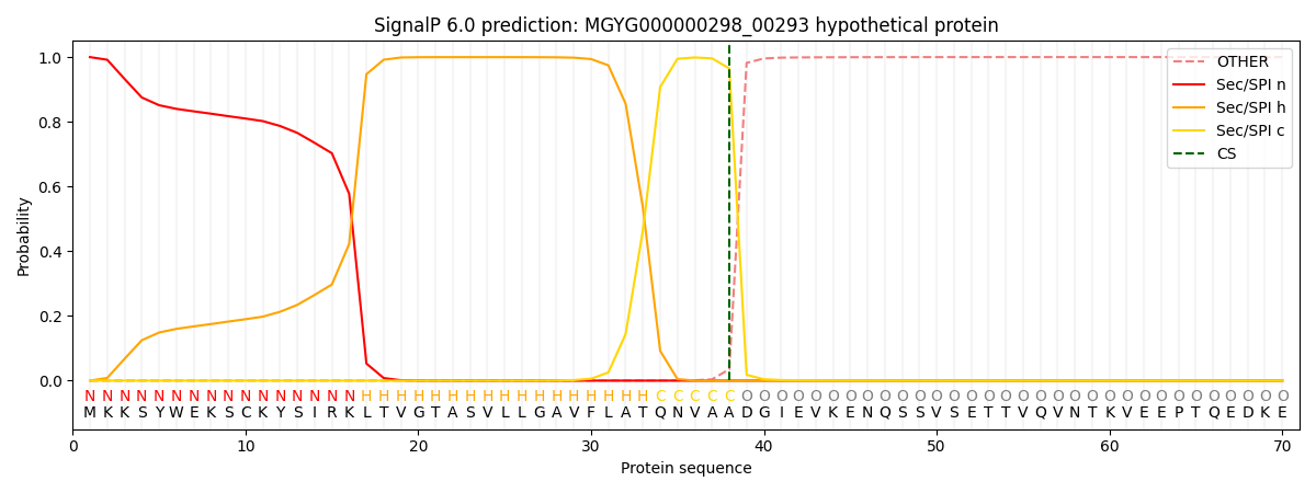
| Other | SP_Sec_SPI | LIPO_Sec_SPII | TAT_Tat_SPI | TATLIP_Sec_SPII | PILIN_Sec_SPIII |
|---|---|---|---|---|---|
| 0.000509 | 0.998722 | 0.000257 | 0.000170 | 0.000161 | 0.000147 |