You are browsing environment: HUMAN GUT
CAZyme Information: MGYG000000330_01183
You are here: Home > Sequence: MGYG000000330_01183
Basic Information |
Genomic context |
Full Sequence |
Enzyme annotations |
CAZy signature domains |
CDD domains |
CAZyme hits |
PDB hits |
Swiss-Prot hits |
SignalP and Lipop annotations |
TMHMM annotations
Basic Information help
| Species | Lentilactobacillus parabuchneri | |||||||||||
|---|---|---|---|---|---|---|---|---|---|---|---|---|
| Lineage | Bacteria; Firmicutes; Bacilli; Lactobacillales; Lactobacillaceae; Lentilactobacillus; Lentilactobacillus parabuchneri | |||||||||||
| CAZyme ID | MGYG000000330_01183 | |||||||||||
| CAZy Family | CBM50 | |||||||||||
| CAZyme Description | hypothetical protein | |||||||||||
| CAZyme Property |
|
|||||||||||
| Genome Property |
|
|||||||||||
| Gene Location | Start: 54938; End: 55489 Strand: + | |||||||||||
CDD Domains download full data without filtering help
| Cdd ID | Domain | E-Value | qStart | qEnd | sStart | sEnd | Domain Description |
|---|---|---|---|---|---|---|---|
| cd00118 | LysM | 1.69e-13 | 7 | 50 | 2 | 45 | Lysin Motif is a small domain involved in binding peptidoglycan. LysM, a small globular domain with approximately 40 amino acids, is a widespread protein module involved in binding peptidoglycan in bacteria and chitin in eukaryotes. The domain was originally identified in enzymes that degrade bacterial cell walls, but proteins involved in many other biological functions also contain this domain. It has been reported that the LysM domain functions as a signal for specific plant-bacteria recognition in bacterial pathogenesis. Many of these enzymes are modular and are composed of catalytic units linked to one or several repeats of LysM domains. LysM domains are found in bacteria and eukaryotes. |
| smart00257 | LysM | 1.10e-10 | 7 | 50 | 1 | 44 | Lysin motif. |
| pfam01476 | LysM | 2.13e-09 | 8 | 50 | 1 | 42 | LysM domain. The LysM (lysin motif) domain is about 40 residues long. It is found in a variety of enzymes involved in bacterial cell wall degradation. This domain may have a general peptidoglycan binding function. The structure of this domain is known. |
| cd14667 | 3D_containing_proteins | 7.17e-09 | 120 | 183 | 4 | 72 | Non-mltA associated 3D domain containing proteins, named for 3 conserved aspartate residues. This family contains the 3D domain, named for its 3 conserved aspartates, including similar uncharacterized proteins. These proteins contain the critical active site aspartate of mltA-like lytic transglycosylases where the 3D domain forms a larger domain with the N-terminal region. This domain is also found in conjunction with numerous other domains such as the Escherichia coli MltA, a membrane-bound lytic transglycosylase comprised of 2 domains separated by a large groove, where the peptidoglycan strand binds. Domain A is made up of an N-terminal and a C-terminal portion, corresponding to the 3D domain and Domain B is inserted within the linear sequence of domain A. MltA is distinct from other bacterial LTs, which are similar to each other. Escherichia coli peptidoglycan lytic transglycosylase (LT) initiates cell wall recycling in response to damage, during bacterial fission, and cleaves peptidoglycan (PG) to create functional spaces in its wall. PG chains (also known as murein), the major components of the bacterial cell wall, are comprised of alternating beta-1-4-linked N-acetylmuramic acid (MurNAc) and N-acetyl-D-glucosamine (GlcNAc), and lytic transglycosylases cleave this beta-1-4 bond. |
| PRK06347 | PRK06347 | 5.34e-07 | 3 | 79 | 328 | 403 | 1,4-beta-N-acetylmuramoylhydrolase. |
CAZyme Hits help
| Hit ID | E-Value | Query Start | Query End | Hit Start | Hit End |
|---|---|---|---|---|---|
| QOP50653.1 | 1.62e-112 | 1 | 183 | 24 | 206 |
| QOJ85509.1 | 1.62e-112 | 1 | 183 | 24 | 206 |
| APR06744.1 | 1.62e-112 | 1 | 183 | 24 | 206 |
| QUX04892.1 | 1.42e-97 | 1 | 183 | 24 | 207 |
| AEB72703.1 | 1.42e-97 | 1 | 183 | 24 | 207 |
Swiss-Prot Hits help
SignalP and Lipop Annotations help
This protein is predicted as OTHER
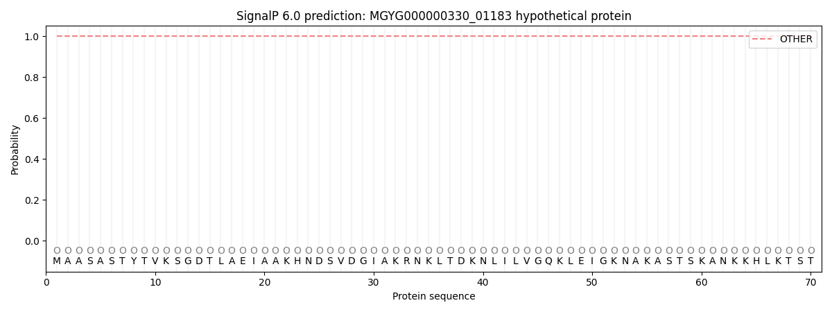
| Other | SP_Sec_SPI | LIPO_Sec_SPII | TAT_Tat_SPI | TATLIP_Sec_SPII | PILIN_Sec_SPIII |
|---|---|---|---|---|---|
| 1.000046 | 0.000009 | 0.000000 | 0.000000 | 0.000000 | 0.000000 |