You are browsing environment: HUMAN GUT
CAZyme Information: MGYG000000442_01057
You are here: Home > Sequence: MGYG000000442_01057
Basic Information |
Genomic context |
Full Sequence |
Enzyme annotations |
CAZy signature domains |
CDD domains |
CAZyme hits |
PDB hits |
Swiss-Prot hits |
SignalP and Lipop annotations |
TMHMM annotations
Basic Information help
| Species | CAG-462 sp003489705 | |||||||||||
|---|---|---|---|---|---|---|---|---|---|---|---|---|
| Lineage | Bacteria; Bacteroidota; Bacteroidia; Bacteroidales; Bacteroidaceae; CAG-462; CAG-462 sp003489705 | |||||||||||
| CAZyme ID | MGYG000000442_01057 | |||||||||||
| CAZy Family | CBM50 | |||||||||||
| CAZyme Description | hypothetical protein | |||||||||||
| CAZyme Property |
|
|||||||||||
| Genome Property |
|
|||||||||||
| Gene Location | Start: 11716; End: 13515 Strand: + | |||||||||||
CDD Domains download full data without filtering help
| Cdd ID | Domain | E-Value | qStart | qEnd | sStart | sEnd | Domain Description |
|---|---|---|---|---|---|---|---|
| cd06268 | PBP1_ABC_transporter_LIVBP-like | 2.53e-12 | 241 | 539 | 1 | 287 | periplasmic binding domain of ATP-binding cassette transporter-like systems that belong to the type 1 periplasmic binding fold protein superfamily. Periplasmic binding domain of ATP-binding cassette transporter-like systems that belong to the type 1 periplasmic binding fold protein superfamily. They are mostly present in archaea and eubacteria, and are primarily involved in scavenging solutes from the environment. ABC-type transporters couple ATP hydrolysis with the uptake and efflux of a wide range of substrates across bacterial membranes, including amino acids, peptides, lipids and sterols, and various drugs. These systems are comprised of transmembrane domains, nucleotide binding domains, and in most bacterial uptake systems, periplasmic binding proteins (PBPs) which transfer the ligand to the extracellular gate of the transmembrane domains. These PBPs bind their substrates selectively and with high affinity. Members of this group include ABC-type Leucine-Isoleucine-Valine-Binding Proteins (LIVBP), which are homologous to the aliphatic amidase transcriptional repressor, AmiC, of Pseudomonas aeruginosa. The uncharacterized periplasmic components of various ABC-type transport systems are included in this group. |
| cd00118 | LysM | 1.25e-10 | 82 | 126 | 1 | 45 | Lysin Motif is a small domain involved in binding peptidoglycan. LysM, a small globular domain with approximately 40 amino acids, is a widespread protein module involved in binding peptidoglycan in bacteria and chitin in eukaryotes. The domain was originally identified in enzymes that degrade bacterial cell walls, but proteins involved in many other biological functions also contain this domain. It has been reported that the LysM domain functions as a signal for specific plant-bacteria recognition in bacterial pathogenesis. Many of these enzymes are modular and are composed of catalytic units linked to one or several repeats of LysM domains. LysM domains are found in bacteria and eukaryotes. |
| cd00118 | LysM | 1.20e-09 | 31 | 72 | 3 | 45 | Lysin Motif is a small domain involved in binding peptidoglycan. LysM, a small globular domain with approximately 40 amino acids, is a widespread protein module involved in binding peptidoglycan in bacteria and chitin in eukaryotes. The domain was originally identified in enzymes that degrade bacterial cell walls, but proteins involved in many other biological functions also contain this domain. It has been reported that the LysM domain functions as a signal for specific plant-bacteria recognition in bacterial pathogenesis. Many of these enzymes are modular and are composed of catalytic units linked to one or several repeats of LysM domains. LysM domains are found in bacteria and eukaryotes. |
| PRK06347 | PRK06347 | 1.45e-09 | 24 | 201 | 401 | 591 | 1,4-beta-N-acetylmuramoylhydrolase. |
| pfam01476 | LysM | 2.93e-09 | 84 | 127 | 1 | 43 | LysM domain. The LysM (lysin motif) domain is about 40 residues long. It is found in a variety of enzymes involved in bacterial cell wall degradation. This domain may have a general peptidoglycan binding function. The structure of this domain is known. |
CAZyme Hits help
| Hit ID | E-Value | Query Start | Query End | Hit Start | Hit End |
|---|---|---|---|---|---|
| ALA74972.1 | 4.63e-227 | 1 | 598 | 1 | 602 |
| QJR68229.1 | 2.65e-226 | 1 | 598 | 1 | 602 |
| QJR60047.1 | 2.65e-226 | 1 | 598 | 1 | 602 |
| AII69224.1 | 2.65e-226 | 1 | 598 | 1 | 602 |
| QJR72564.1 | 2.65e-226 | 1 | 598 | 1 | 602 |
PDB Hits download full data without filtering help
| Hit ID | E-Value | Query Start | Query End | Hit Start | Hit End | Description |
|---|---|---|---|---|---|---|
| 4B8V_A | 1.48e-07 | 25 | 202 | 38 | 217 | ChainA, Extracellular Protein 6 [Fulvia fulva],4B9H_A Chain A, Extracellular Protein 6 [Fulvia fulva] |
Swiss-Prot Hits download full data without filtering help
| Hit ID | E-Value | Query Start | Query End | Hit Start | Hit End | Description |
|---|---|---|---|---|---|---|
| P44493 | 1.73e-10 | 26 | 201 | 289 | 429 | Probable N-acetylmuramoyl-L-alanine amidase AmiB OS=Haemophilus influenzae (strain ATCC 51907 / DSM 11121 / KW20 / Rd) OX=71421 GN=amiB PE=3 SV=1 |
| Q49UX4 | 2.36e-07 | 25 | 185 | 23 | 178 | N-acetylmuramoyl-L-alanine amidase sle1 OS=Staphylococcus saprophyticus subsp. saprophyticus (strain ATCC 15305 / DSM 20229 / NCIMB 8711 / NCTC 7292 / S-41) OX=342451 GN=sle1 PE=3 SV=1 |
| Q9P403 | 1.11e-06 | 17 | 127 | 100 | 228 | Intracellular hyphae protein 1 OS=Colletotrichum lindemuthianum OX=290576 GN=CIH1 PE=1 SV=1 |
| Q2G0D4 | 2.09e-06 | 31 | 110 | 29 | 117 | Probable autolysin SsaALP OS=Staphylococcus aureus (strain NCTC 8325 / PS 47) OX=93061 GN=SAOUHSC_00671 PE=1 SV=1 |
| O05495 | 2.37e-06 | 29 | 128 | 2 | 94 | Putative sporulation-specific glycosylase YdhD OS=Bacillus subtilis (strain 168) OX=224308 GN=ydhD PE=1 SV=2 |
SignalP and Lipop Annotations help
This protein is predicted as SP
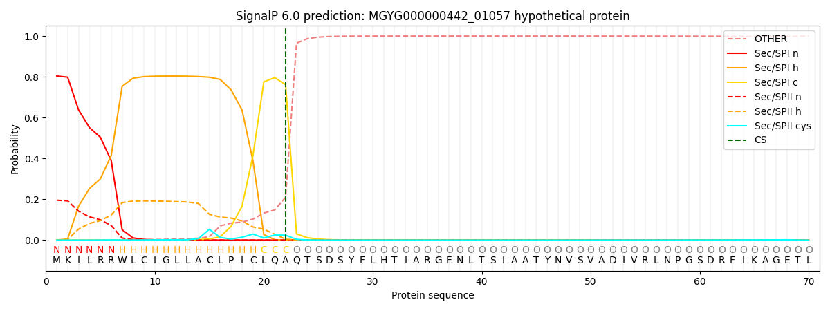
| Other | SP_Sec_SPI | LIPO_Sec_SPII | TAT_Tat_SPI | TATLIP_Sec_SPII | PILIN_Sec_SPIII |
|---|---|---|---|---|---|
| 0.001144 | 0.793138 | 0.204810 | 0.000329 | 0.000296 | 0.000261 |