You are browsing environment: HUMAN GUT
CAZyme Information: MGYG000000555_00922
You are here: Home > Sequence: MGYG000000555_00922
Basic Information |
Genomic context |
Full Sequence |
Enzyme annotations |
CAZy signature domains |
CDD domains |
CAZyme hits |
PDB hits |
Swiss-Prot hits |
SignalP and Lipop annotations |
TMHMM annotations
Basic Information help
| Species | Cellulosilyticum sp900556665 | |||||||||||
|---|---|---|---|---|---|---|---|---|---|---|---|---|
| Lineage | Bacteria; Firmicutes_A; Clostridia; Lachnospirales; Cellulosilyticaceae; Cellulosilyticum; Cellulosilyticum sp900556665 | |||||||||||
| CAZyme ID | MGYG000000555_00922 | |||||||||||
| CAZy Family | CBM65 | |||||||||||
| CAZyme Description | hypothetical protein | |||||||||||
| CAZyme Property |
|
|||||||||||
| Genome Property |
|
|||||||||||
| Gene Location | Start: 34; End: 1563 Strand: + | |||||||||||
CAZyme Signature Domains help
| Family | Start | End | Evalue | family coverage |
|---|---|---|---|---|
| CBM65 | 101 | 215 | 3.3e-38 | 0.9649122807017544 |
| CBM65 | 1 | 91 | 3.2e-33 | 0.7631578947368421 |
| CBM2 | 310 | 401 | 8.8e-23 | 0.9108910891089109 |
| CBM2 | 416 | 499 | 1.8e-21 | 0.8316831683168316 |
CDD Domains download full data without filtering help
| Cdd ID | Domain | E-Value | qStart | qEnd | sStart | sEnd | Domain Description |
|---|---|---|---|---|---|---|---|
| pfam18259 | CBM65_1 | 4.86e-22 | 105 | 215 | 5 | 113 | Carbohydrate binding module 65 domain 1. This domain is found in the non-catalytic carbohydrate binding module 65B (CMB65B) present in Eubacterium cellulosolvens. CBMs are present in plant cell wall degrading enzymes and are responsible for targeting, which enhances catalysis. CBM65s display higher affinity for oligosaccharides, such as cellohexaose, and particularly polysaccharides than cellotetraose, which fully occupies the core component of the substrate binding cleft. The concave surface presented by beta-sheet 2 comprises the beta-glucan binding site in CBM65s. C6 of all the backbone glucose moieties makes extensive hydrophobic interactions with the surface tryptophans of CBM65s. Three out of the four surface Trp are highly conserved. The conserved metal ion site typical of CBMs is absent in this CBM65 family. |
| pfam18259 | CBM65_1 | 7.11e-19 | 4 | 90 | 29 | 113 | Carbohydrate binding module 65 domain 1. This domain is found in the non-catalytic carbohydrate binding module 65B (CMB65B) present in Eubacterium cellulosolvens. CBMs are present in plant cell wall degrading enzymes and are responsible for targeting, which enhances catalysis. CBM65s display higher affinity for oligosaccharides, such as cellohexaose, and particularly polysaccharides than cellotetraose, which fully occupies the core component of the substrate binding cleft. The concave surface presented by beta-sheet 2 comprises the beta-glucan binding site in CBM65s. C6 of all the backbone glucose moieties makes extensive hydrophobic interactions with the surface tryptophans of CBM65s. Three out of the four surface Trp are highly conserved. The conserved metal ion site typical of CBMs is absent in this CBM65 family. |
| smart00637 | CBD_II | 2.09e-18 | 315 | 401 | 1 | 87 | CBD_II domain. |
| pfam00553 | CBM_2 | 5.30e-17 | 420 | 499 | 7 | 87 | Cellulose binding domain. Two tryptophan residues are involved in cellulose binding. Cellulose binding domain found in bacteria. |
| smart00637 | CBD_II | 7.03e-17 | 422 | 508 | 2 | 89 | CBD_II domain. |
CAZyme Hits help
| Hit ID | E-Value | Query Start | Query End | Hit Start | Hit End |
|---|---|---|---|---|---|
| ACZ98591.1 | 0.0 | 1 | 509 | 747 | 1255 |
| ADZ83488.1 | 2.71e-75 | 94 | 495 | 847 | 1163 |
| QEH68935.1 | 6.32e-74 | 94 | 495 | 847 | 1163 |
| QEH69636.1 | 2.46e-41 | 308 | 494 | 63 | 249 |
| ADZ83921.1 | 6.97e-41 | 308 | 509 | 63 | 266 |
PDB Hits download full data without filtering help
| Hit ID | E-Value | Query Start | Query End | Hit Start | Hit End | Description |
|---|---|---|---|---|---|---|
| 6BT9_A | 2.23e-18 | 313 | 385 | 582 | 654 | ChitinaseChiA74 from Bacillus thuringiensis [Bacillus thuringiensis],6BT9_B Chitinase ChiA74 from Bacillus thuringiensis [Bacillus thuringiensis] |
| 2RTT_A | 8.52e-13 | 316 | 401 | 12 | 95 | Solutionstructure of the chitin-binding domain of Chi18aC from Streptomyces coelicolor [Streptomyces coelicolor] |
| 1EXG_A | 9.10e-10 | 316 | 400 | 15 | 100 | ChainA, EXO-1,4-BETA-D-GLYCANASE [Cellulomonas fimi],1EXH_A Chain A, EXO-1,4-BETA-D-GLYCANASE [Cellulomonas fimi] |
| 6QFS_A | 1.54e-09 | 316 | 400 | 11 | 96 | ChainA, Exoglucanase/xylanase [Cellulomonas fimi],6QFS_B Chain B, Exoglucanase/xylanase [Cellulomonas fimi],6QFS_C Chain C, Exoglucanase/xylanase [Cellulomonas fimi],6QFS_D Chain D, Exoglucanase/xylanase [Cellulomonas fimi],6QFS_E Chain E, Exoglucanase/xylanase [Cellulomonas fimi],6QFS_F Chain F, Exoglucanase/xylanase [Cellulomonas fimi],6QFS_G Chain G, Exoglucanase/xylanase [Cellulomonas fimi],6QFS_H Chain H, Exoglucanase/xylanase [Cellulomonas fimi] |
| 6F7E_A | 3.29e-08 | 313 | 393 | 10 | 92 | NMRsolution structure of the cellulose-binding family 2 carbohydrate binding domain (CBM2) from ScLPMO9C [Streptomyces coelicolor A3(2)] |
Swiss-Prot Hits download full data without filtering help
| Hit ID | E-Value | Query Start | Query End | Hit Start | Hit End | Description |
|---|---|---|---|---|---|---|
| P36909 | 8.00e-11 | 316 | 417 | 42 | 141 | Chitinase C OS=Streptomyces lividans OX=1916 GN=chiC PE=2 SV=1 |
| P64906 | 8.82e-10 | 310 | 399 | 42 | 132 | Uncharacterized protein Mb2009 OS=Mycobacterium bovis (strain ATCC BAA-935 / AF2122/97) OX=233413 GN=BQ2027_MB2009 PE=3 SV=1 |
| P9WLQ0 | 8.82e-10 | 310 | 399 | 42 | 132 | Uncharacterized protein MT2041 OS=Mycobacterium tuberculosis (strain CDC 1551 / Oshkosh) OX=83331 GN=MT2041 PE=3 SV=1 |
| P9WLQ1 | 8.82e-10 | 310 | 399 | 42 | 132 | Uncharacterized protein Rv1987 OS=Mycobacterium tuberculosis (strain ATCC 25618 / H37Rv) OX=83332 GN=Rv1987 PE=1 SV=1 |
| P50899 | 5.06e-09 | 422 | 499 | 997 | 1075 | Exoglucanase B OS=Cellulomonas fimi (strain ATCC 484 / DSM 20113 / JCM 1341 / NBRC 15513 / NCIMB 8980 / NCTC 7547) OX=590998 GN=cbhB PE=1 SV=1 |
SignalP and Lipop Annotations help
This protein is predicted as OTHER
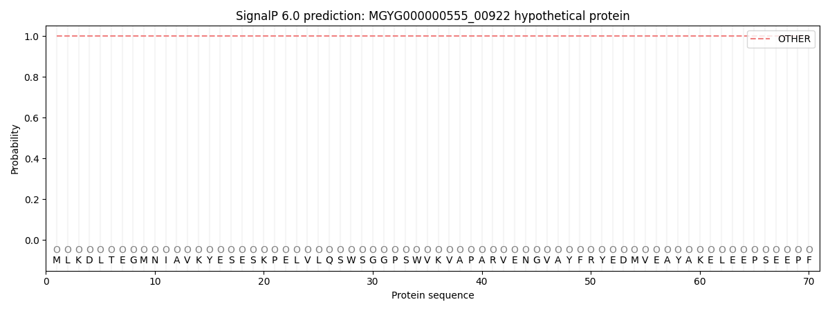
| Other | SP_Sec_SPI | LIPO_Sec_SPII | TAT_Tat_SPI | TATLIP_Sec_SPII | PILIN_Sec_SPIII |
|---|---|---|---|---|---|
| 1.000073 | 0.000000 | 0.000000 | 0.000000 | 0.000000 | 0.000000 |