You are browsing environment: HUMAN GUT
CAZyme Information: MGYG000000584_01702
You are here: Home > Sequence: MGYG000000584_01702
Basic Information |
Genomic context |
Full Sequence |
Enzyme annotations |
CAZy signature domains |
CDD domains |
CAZyme hits |
PDB hits |
Swiss-Prot hits |
SignalP and Lipop annotations |
TMHMM annotations
Basic Information help
| Species | RC9 sp900544195 | |||||||||||
|---|---|---|---|---|---|---|---|---|---|---|---|---|
| Lineage | Bacteria; Bacteroidota; Bacteroidia; Bacteroidales; UBA932; RC9; RC9 sp900544195 | |||||||||||
| CAZyme ID | MGYG000000584_01702 | |||||||||||
| CAZy Family | GT0 | |||||||||||
| CAZyme Description | UDP-N-acetylglucosamine 2-epimerase | |||||||||||
| CAZyme Property |
|
|||||||||||
| Genome Property |
|
|||||||||||
| Gene Location | Start: 21943; End: 23109 Strand: + | |||||||||||
CDD Domains download full data without filtering help
| Cdd ID | Domain | E-Value | qStart | qEnd | sStart | sEnd | Domain Description |
|---|---|---|---|---|---|---|---|
| COG0381 | WecB | 1.90e-168 | 1 | 378 | 7 | 369 | UDP-N-acetylglucosamine 2-epimerase [Cell wall/membrane/envelope biogenesis]. |
| TIGR00236 | wecB | 5.39e-158 | 1 | 377 | 4 | 361 | UDP-N-acetylglucosamine 2-epimerase. This cytosolic enzyme converts UDP-N-acetyl-D-glucosamine to UDP-N-acetyl-D-mannosamine. In E. coli, this is the first step in the pathway of enterobacterial common antigen biosynthesis.Members of this orthology group have many gene symbols, often reflecting the overall activity of the pathway and/or operon that includes it. Symbols include epsC (exopolysaccharide C) in Burkholderia solanacerum, cap8P (type 8 capsule P) in Staphylococcus aureus, and nfrC in an older designation based on the effects of deletion on phage N4 adsorption. Epimerase activity was also demonstrated in a bifunctional rat enzyme, for which the N-terminal domain appears to be orthologous. The set of proteins found above the suggested cutoff includes E. coli WecB in one of two deeply branched clusters and the rat UDP-N-acetylglucosamine 2-epimerase domain in the other. [Cell envelope, Biosynthesis and degradation of surface polysaccharides and lipopolysaccharides] |
| cd03786 | GTB_UDP-GlcNAc_2-Epimerase | 8.54e-140 | 1 | 377 | 3 | 364 | UDP-N-acetylglucosamine 2-epimerase and similar proteins. Bacterial members of the UDP-N-Acetylglucosamine (GlcNAc) 2-Epimerase family (EC 5.1.3.14) are known to catalyze the reversible interconversion of UDP-GlcNAc and UDP-N-acetylmannosamine (UDP-ManNAc). The enzyme serves to produce an activated form of ManNAc residues (UDP-ManNAc) for use in the biosynthesis of a variety of cell surface polysaccharides; The mammalian enzyme is bifunctional, catalyzing both the inversion of stereochemistry at C-2 and the hydrolysis of the UDP-sugar linkage to generate free ManNAc. It also catalyzes the phosphorylation of ManNAc to generate ManNAc 6-phosphate, a precursor to salic acids. In mammals, sialic acids are found at the termini of oligosaccharides in a large variety of cell surface glycoconjugates and are key mediators of cell-cell recognition events. Mutations in human members of this family have been associated with Sialuria, a rare disease caused by the disorders of sialic acid metabolism. This family belongs to the GT-B structural superfamily of glycoslytransferases, which have characteristic N- and C-terminal domains each containing a typical Rossmann fold. The two domains have high structural homology despite minimal sequence homology. The large cleft that separates the two domains includes the catalytic center and permits a high degree of flexibility. |
| pfam02350 | Epimerase_2 | 1.42e-134 | 18 | 377 | 1 | 335 | UDP-N-acetylglucosamine 2-epimerase. This family consists of UDP-N-acetylglucosamine 2-epimerases EC:5.1.3.14 this enzyme catalyzes the production of UDP-ManNAc from UDP-GlcNAc. Note that some of the enzymes is this family are bifunctional, in these instances Pfam matches only the N-terminal half of the protein suggesting that the additional C-terminal part (when compared to mono-functional members of this family) is responsible for the UPD-N-acetylmannosamine kinase activity of these enzymes. This hypothesis is further supported by the assumption that the C-terminal part of rat Gne is the kinase domain. |
| COG0438 | RfaB | 0.003 | 204 | 360 | 194 | 352 | Glycosyltransferase involved in cell wall bisynthesis [Cell wall/membrane/envelope biogenesis]. |
CAZyme Hits help
| Hit ID | E-Value | Query Start | Query End | Hit Start | Hit End |
|---|---|---|---|---|---|
| QQN33861.1 | 5.43e-150 | 1 | 384 | 4 | 376 |
| AZP48621.1 | 3.12e-149 | 1 | 384 | 4 | 376 |
| AZP44285.1 | 3.12e-149 | 1 | 384 | 4 | 376 |
| QBJ09996.1 | 3.12e-149 | 1 | 384 | 4 | 376 |
| AZP53030.1 | 8.89e-149 | 1 | 384 | 4 | 376 |
PDB Hits download full data without filtering help
| Hit ID | E-Value | Query Start | Query End | Hit Start | Hit End | Description |
|---|---|---|---|---|---|---|
| 3DZC_A | 8.06e-146 | 1 | 377 | 29 | 394 | 2.35Angstrom resolution structure of WecB (VC0917), a UDP-N-acetylglucosamine 2-epimerase from Vibrio cholerae. [Vibrio cholerae],3DZC_B 2.35 Angstrom resolution structure of WecB (VC0917), a UDP-N-acetylglucosamine 2-epimerase from Vibrio cholerae. [Vibrio cholerae] |
| 1F6D_A | 5.39e-144 | 1 | 382 | 4 | 374 | TheStructure Of Udp-N-Acetylglucosamine 2-Epimerase From E. Coli. [Escherichia coli],1F6D_B The Structure Of Udp-N-Acetylglucosamine 2-Epimerase From E. Coli. [Escherichia coli],1F6D_C The Structure Of Udp-N-Acetylglucosamine 2-Epimerase From E. Coli. [Escherichia coli],1F6D_D The Structure Of Udp-N-Acetylglucosamine 2-Epimerase From E. Coli. [Escherichia coli] |
| 1VGV_A | 7.10e-144 | 1 | 382 | 4 | 374 | Crystalstructure of UDP-N-acetylglucosamine_2 epimerase [Escherichia coli],1VGV_B Crystal structure of UDP-N-acetylglucosamine_2 epimerase [Escherichia coli],1VGV_C Crystal structure of UDP-N-acetylglucosamine_2 epimerase [Escherichia coli],1VGV_D Crystal structure of UDP-N-acetylglucosamine_2 epimerase [Escherichia coli] |
| 5DLD_A | 4.40e-142 | 1 | 377 | 13 | 378 | CrystalStructure of a UDP-N-acetylglucosamine 2-epimerase from Burkholderia vietnamiensis complexed with UDP-GlcNAc and UDP [Burkholderia vietnamiensis G4] |
| 6VLB_A | 1.55e-134 | 1 | 377 | 4 | 368 | Crystalstructure of ligand-free UDP-GlcNAc 2-epimerase from Neisseria meningitidis [Neisseria meningitidis Z2491],6VLB_B Crystal structure of ligand-free UDP-GlcNAc 2-epimerase from Neisseria meningitidis [Neisseria meningitidis Z2491],6VLC_A Crystal structure of UDP-GlcNAc 2-epimerase from Neisseria meningitidis bound to UDP-GlcNAc [Neisseria meningitidis Z2491],6VLC_B Crystal structure of UDP-GlcNAc 2-epimerase from Neisseria meningitidis bound to UDP-GlcNAc [Neisseria meningitidis Z2491],6VLC_C Crystal structure of UDP-GlcNAc 2-epimerase from Neisseria meningitidis bound to UDP-GlcNAc [Neisseria meningitidis Z2491],6VLC_D Crystal structure of UDP-GlcNAc 2-epimerase from Neisseria meningitidis bound to UDP-GlcNAc [Neisseria meningitidis Z2491] |
Swiss-Prot Hits download full data without filtering help
| Hit ID | E-Value | Query Start | Query End | Hit Start | Hit End | Description |
|---|---|---|---|---|---|---|
| P52641 | 7.40e-151 | 1 | 377 | 5 | 370 | Probable UDP-N-acetylglucosamine 2-epimerase OS=Ralstonia solanacearum OX=305 GN=epsC PE=3 SV=2 |
| P58600 | 2.11e-150 | 1 | 377 | 5 | 370 | Probable UDP-N-acetylglucosamine 2-epimerase OS=Ralstonia solanacearum (strain GMI1000) OX=267608 GN=epsC PE=3 SV=1 |
| Q8XAR8 | 4.40e-150 | 1 | 382 | 4 | 374 | UDP-N-acetylglucosamine 2-epimerase OS=Escherichia coli O157:H7 OX=83334 GN=wecB PE=3 SV=1 |
| Q8ZAE3 | 1.78e-149 | 1 | 382 | 4 | 374 | UDP-N-acetylglucosamine 2-epimerase OS=Yersinia pestis OX=632 GN=wecB PE=3 SV=1 |
| P27828 | 3.58e-149 | 1 | 382 | 4 | 374 | UDP-N-acetylglucosamine 2-epimerase OS=Escherichia coli (strain K12) OX=83333 GN=wecB PE=1 SV=2 |
SignalP and Lipop Annotations help
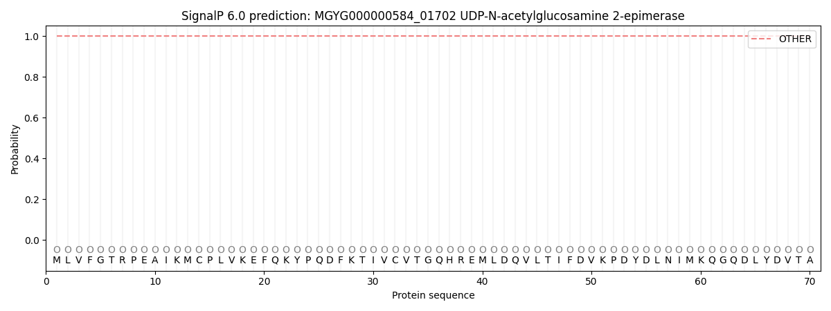
This protein is predicted as OTHER
| Other | SP_Sec_SPI | LIPO_Sec_SPII | TAT_Tat_SPI | TATLIP_Sec_SPII | PILIN_Sec_SPIII |
|---|---|---|---|---|---|
| 1.000039 | 0.000000 | 0.000000 | 0.000000 | 0.000000 | 0.000000 |