You are browsing environment: HUMAN GUT
CAZyme Information: MGYG000000624_00913
You are here: Home > Sequence: MGYG000000624_00913
Basic Information |
Genomic context |
Full Sequence |
Enzyme annotations |
CAZy signature domains |
CDD domains |
CAZyme hits |
PDB hits |
Swiss-Prot hits |
SignalP and Lipop annotations |
TMHMM annotations
Basic Information help
| Species | ||||||||||||
|---|---|---|---|---|---|---|---|---|---|---|---|---|
| Lineage | Bacteria; Proteobacteria; Gammaproteobacteria; Burkholderiales; Burkholderiaceae; Duodenibacillus; | |||||||||||
| CAZyme ID | MGYG000000624_00913 | |||||||||||
| CAZy Family | GH8 | |||||||||||
| CAZyme Description | Endoglucanase | |||||||||||
| CAZyme Property |
|
|||||||||||
| Genome Property |
|
|||||||||||
| Gene Location | Start: 6670; End: 7983 Strand: - | |||||||||||
CDD Domains download full data without filtering help
| Cdd ID | Domain | E-Value | qStart | qEnd | sStart | sEnd | Domain Description |
|---|---|---|---|---|---|---|---|
| PRK11097 | PRK11097 | 2.07e-153 | 6 | 360 | 4 | 367 | cellulase. |
| COG3405 | BcsZ | 4.70e-87 | 2 | 351 | 1 | 350 | Endo-1,4-beta-D-glucanase Y [Carbohydrate transport and metabolism]. |
| pfam01270 | Glyco_hydro_8 | 9.32e-63 | 30 | 347 | 8 | 321 | Glycosyl hydrolases family 8. |
| NF033839 | PspC_subgroup_2 | 0.002 | 368 | 429 | 314 | 371 | pneumococcal surface protein PspC, LPXTG-anchored form. The pneumococcal surface protein PspC, as described in Streptococcus pneumoniae, is a repetitive and highly variable protein, recognized by a conserved N-terminal domain and also by genomic location. This form, subgroup 2, is anchored covalently after cleavage by sortase at a C-terminal LPXTG site. The other form, subgroup 1, has variable numbers of a choline-binding repeat in the C-terminal region, and is also known as choline-binding protein A. |
CAZyme Hits help
| Hit ID | E-Value | Query Start | Query End | Hit Start | Hit End |
|---|---|---|---|---|---|
| QDA53874.1 | 7.88e-143 | 14 | 359 | 6 | 360 |
| QQS88762.1 | 8.16e-143 | 4 | 361 | 3 | 363 |
| ANU67169.1 | 1.35e-120 | 7 | 361 | 3 | 352 |
| QQQ96024.1 | 1.35e-120 | 7 | 361 | 3 | 352 |
| QNT42998.1 | 5.05e-103 | 10 | 357 | 9 | 361 |
PDB Hits download full data without filtering help
| Hit ID | E-Value | Query Start | Query End | Hit Start | Hit End | Description |
|---|---|---|---|---|---|---|
| 4Q2B_A | 1.20e-99 | 24 | 357 | 1 | 339 | Thecrystal structure of an endo-1,4-D-glucanase from Pseudomonas putida KT2440 [Pseudomonas putida KT2440],4Q2B_B The crystal structure of an endo-1,4-D-glucanase from Pseudomonas putida KT2440 [Pseudomonas putida KT2440],4Q2B_C The crystal structure of an endo-1,4-D-glucanase from Pseudomonas putida KT2440 [Pseudomonas putida KT2440],4Q2B_D The crystal structure of an endo-1,4-D-glucanase from Pseudomonas putida KT2440 [Pseudomonas putida KT2440],4Q2B_E The crystal structure of an endo-1,4-D-glucanase from Pseudomonas putida KT2440 [Pseudomonas putida KT2440],4Q2B_F The crystal structure of an endo-1,4-D-glucanase from Pseudomonas putida KT2440 [Pseudomonas putida KT2440] |
| 7F81_A | 2.04e-98 | 24 | 358 | 5 | 340 | ChainA, Glucanase [Enterobacter sp. CJF-002],7F81_B Chain B, Glucanase [Enterobacter sp. CJF-002],7F81_C Chain C, Glucanase [Enterobacter sp. CJF-002],7F81_D Chain D, Glucanase [Enterobacter sp. CJF-002] |
| 7F82_A | 5.77e-98 | 24 | 358 | 5 | 340 | ChainA, Glucanase [Enterobacter sp. CJF-002],7F82_B Chain B, Glucanase [Enterobacter sp. CJF-002],7F82_C Chain C, Glucanase [Enterobacter sp. CJF-002],7F82_D Chain D, Glucanase [Enterobacter sp. CJF-002] |
| 3QXQ_A | 6.66e-95 | 24 | 357 | 1 | 335 | Structureof the bacterial cellulose synthase subunit Z in complex with cellopentaose [Escherichia coli K-12],3QXQ_B Structure of the bacterial cellulose synthase subunit Z in complex with cellopentaose [Escherichia coli K-12],3QXQ_C Structure of the bacterial cellulose synthase subunit Z in complex with cellopentaose [Escherichia coli K-12],3QXQ_D Structure of the bacterial cellulose synthase subunit Z in complex with cellopentaose [Escherichia coli K-12] |
| 3QXF_A | 6.02e-90 | 24 | 357 | 1 | 335 | Structureof the bacterial cellulose synthase subunit Z [Escherichia coli K-12],3QXF_B Structure of the bacterial cellulose synthase subunit Z [Escherichia coli K-12],3QXF_C Structure of the bacterial cellulose synthase subunit Z [Escherichia coli K-12],3QXF_D Structure of the bacterial cellulose synthase subunit Z [Escherichia coli K-12] |
Swiss-Prot Hits download full data without filtering help
| Hit ID | E-Value | Query Start | Query End | Hit Start | Hit End | Description |
|---|---|---|---|---|---|---|
| Q8X5L9 | 1.36e-97 | 4 | 357 | 2 | 356 | Endoglucanase OS=Escherichia coli O157:H7 OX=83334 GN=bcsZ PE=3 SV=1 |
| Q8Z289 | 2.80e-97 | 6 | 357 | 5 | 357 | Endoglucanase OS=Salmonella typhi OX=90370 GN=bcsZ PE=3 SV=1 |
| Q8ZLB7 | 5.60e-97 | 6 | 357 | 5 | 357 | Endoglucanase OS=Salmonella typhimurium (strain LT2 / SGSC1412 / ATCC 700720) OX=99287 GN=bcsZ PE=3 SV=1 |
| P37651 | 3.06e-96 | 4 | 357 | 2 | 356 | Endoglucanase OS=Escherichia coli (strain K12) OX=83333 GN=bcsZ PE=1 SV=1 |
| P58935 | 1.93e-80 | 11 | 357 | 15 | 371 | Endoglucanase OS=Xanthomonas axonopodis pv. citri (strain 306) OX=190486 GN=bcsZ PE=3 SV=1 |
SignalP and Lipop Annotations help
This protein is predicted as SP
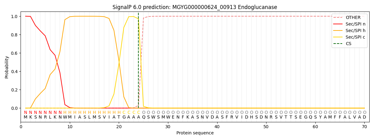
| Other | SP_Sec_SPI | LIPO_Sec_SPII | TAT_Tat_SPI | TATLIP_Sec_SPII | PILIN_Sec_SPIII |
|---|---|---|---|---|---|
| 0.000654 | 0.998344 | 0.000257 | 0.000262 | 0.000239 | 0.000216 |