You are browsing environment: HUMAN GUT
CAZyme Information: MGYG000000711_01559
You are here: Home > Sequence: MGYG000000711_01559
Basic Information |
Genomic context |
Full Sequence |
Enzyme annotations |
CAZy signature domains |
CDD domains |
CAZyme hits |
PDB hits |
Swiss-Prot hits |
SignalP and Lipop annotations |
TMHMM annotations
Basic Information help
| Species | ||||||||||||
|---|---|---|---|---|---|---|---|---|---|---|---|---|
| Lineage | Bacteria; Firmicutes_A; Clostridia; Peptostreptococcales; Anaerovoracaceae; CAG-238; | |||||||||||
| CAZyme ID | MGYG000000711_01559 | |||||||||||
| CAZy Family | CBM50 | |||||||||||
| CAZyme Description | hypothetical protein | |||||||||||
| CAZyme Property |
|
|||||||||||
| Genome Property |
|
|||||||||||
| Gene Location | Start: 1656; End: 2144 Strand: - | |||||||||||
CDD Domains download full data without filtering help
| Cdd ID | Domain | E-Value | qStart | qEnd | sStart | sEnd | Domain Description |
|---|---|---|---|---|---|---|---|
| cd00118 | LysM | 9.55e-20 | 113 | 157 | 1 | 45 | Lysin Motif is a small domain involved in binding peptidoglycan. LysM, a small globular domain with approximately 40 amino acids, is a widespread protein module involved in binding peptidoglycan in bacteria and chitin in eukaryotes. The domain was originally identified in enzymes that degrade bacterial cell walls, but proteins involved in many other biological functions also contain this domain. It has been reported that the LysM domain functions as a signal for specific plant-bacteria recognition in bacterial pathogenesis. Many of these enzymes are modular and are composed of catalytic units linked to one or several repeats of LysM domains. LysM domains are found in bacteria and eukaryotes. |
| smart00257 | LysM | 1.05e-18 | 114 | 157 | 1 | 44 | Lysin motif. |
| pfam01476 | LysM | 2.31e-17 | 115 | 157 | 1 | 42 | LysM domain. The LysM (lysin motif) domain is about 40 residues long. It is found in a variety of enzymes involved in bacterial cell wall degradation. This domain may have a general peptidoglycan binding function. The structure of this domain is known. |
| TIGR02899 | spore_safA | 2.19e-14 | 117 | 157 | 1 | 42 | spore coat assembly protein SafA. SafA (YrbB) (SafA) of Bacillus subtilis is a protein found at the interface of the spore cortex and spore coat, and is dependent on SpoVID for its localization. This model is based on the N-terminal LysM (lysin motif) domain (see pfamAM model pfam01476) of SafA and, from several other spore-forming species, the protein with the most similar N-terminal region. However, this set of proteins differs greatly in C-terminal of the LysM domaim; blocks of 12-residue and 13-residue repeats are found in the Bacillus cereus group, tandem LysM domains in Thermoanaerobacter tengcongensis MB4, etc. in which one of which is found in most examples of endospore-forming bacteria. [Cellular processes, Sporulation and germination] |
| COG1388 | LysM | 1.64e-12 | 82 | 159 | 38 | 112 | LysM repeat [Cell wall/membrane/envelope biogenesis]. |
CAZyme Hits help
| Hit ID | E-Value | Query Start | Query End | Hit Start | Hit End |
|---|---|---|---|---|---|
| AMJ42415.1 | 3.37e-46 | 1 | 161 | 355 | 519 |
| ALX07044.1 | 3.23e-24 | 3 | 161 | 358 | 521 |
| ANV74780.1 | 3.23e-24 | 3 | 161 | 358 | 521 |
| QCU03902.1 | 2.09e-23 | 1 | 161 | 345 | 510 |
| AEY64317.1 | 1.03e-22 | 3 | 160 | 358 | 520 |
PDB Hits download full data without filtering help
| Hit ID | E-Value | Query Start | Query End | Hit Start | Hit End | Description |
|---|---|---|---|---|---|---|
| 4UZ2_A | 3.28e-07 | 111 | 155 | 1 | 44 | Crystalstructure of the N-terminal LysM domains from the putative NlpC/P60 D,L endopeptidase from T. thermophilus [Thermus thermophilus HB8],4UZ2_B Crystal structure of the N-terminal LysM domains from the putative NlpC/P60 D,L endopeptidase from T. thermophilus [Thermus thermophilus HB8],4UZ2_C Crystal structure of the N-terminal LysM domains from the putative NlpC/P60 D,L endopeptidase from T. thermophilus [Thermus thermophilus HB8],4UZ2_D Crystal structure of the N-terminal LysM domains from the putative NlpC/P60 D,L endopeptidase from T. thermophilus [Thermus thermophilus HB8],4UZ3_A Crystal structure of the N-terminal LysM domains from the putative NlpC/P60 D,L endopeptidase from T. thermophilus bound to N-acetyl-chitohexaose [Thermus thermophilus HB8],4UZ3_B Crystal structure of the N-terminal LysM domains from the putative NlpC/P60 D,L endopeptidase from T. thermophilus bound to N-acetyl-chitohexaose [Thermus thermophilus HB8],4UZ3_C Crystal structure of the N-terminal LysM domains from the putative NlpC/P60 D,L endopeptidase from T. thermophilus bound to N-acetyl-chitohexaose [Thermus thermophilus HB8] |
Swiss-Prot Hits download full data without filtering help
| Hit ID | E-Value | Query Start | Query End | Hit Start | Hit End | Description |
|---|---|---|---|---|---|---|
| O32062 | 8.39e-07 | 114 | 155 | 3 | 45 | SpoIVD-associated factor A OS=Bacillus subtilis (strain 168) OX=224308 GN=safA PE=1 SV=1 |
| P37531 | 1.18e-06 | 114 | 157 | 3 | 46 | Cortical fragment-lytic enzyme OS=Bacillus subtilis (strain 168) OX=224308 GN=sleL PE=1 SV=2 |
| Q6B4J5 | 3.20e-06 | 114 | 162 | 3 | 53 | Spore coat assembly protein ExsA OS=Bacillus cereus OX=1396 GN=exsA PE=2 SV=1 |
| Q5HGI5 | 3.85e-06 | 114 | 155 | 176 | 217 | Probable cell wall hydrolase LytN OS=Staphylococcus aureus (strain COL) OX=93062 GN=lytN PE=3 SV=1 |
| Q6G9W6 | 3.85e-06 | 114 | 155 | 176 | 217 | Probable cell wall hydrolase LytN OS=Staphylococcus aureus (strain MSSA476) OX=282459 GN=lytN PE=3 SV=2 |
SignalP and Lipop Annotations help
This protein is predicted as OTHER
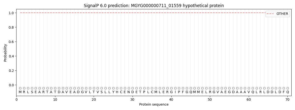
| Other | SP_Sec_SPI | LIPO_Sec_SPII | TAT_Tat_SPI | TATLIP_Sec_SPII | PILIN_Sec_SPIII |
|---|---|---|---|---|---|
| 1.000016 | 0.000016 | 0.000000 | 0.000000 | 0.000000 | 0.000000 |