You are browsing environment: HUMAN GUT
CAZyme Information: MGYG000000730_01646
You are here: Home > Sequence: MGYG000000730_01646
Basic Information |
Genomic context |
Full Sequence |
Enzyme annotations |
CAZy signature domains |
CDD domains |
CAZyme hits |
PDB hits |
Swiss-Prot hits |
SignalP and Lipop annotations |
TMHMM annotations
Basic Information help
| Species | Phil1 sp001940855 | |||||||||||
|---|---|---|---|---|---|---|---|---|---|---|---|---|
| Lineage | Bacteria; Firmicutes_A; Clostridia_A; Christensenellales; CAG-138; Phil1; Phil1 sp001940855 | |||||||||||
| CAZyme ID | MGYG000000730_01646 | |||||||||||
| CAZy Family | CBM50 | |||||||||||
| CAZyme Description | hypothetical protein | |||||||||||
| CAZyme Property |
|
|||||||||||
| Genome Property |
|
|||||||||||
| Gene Location | Start: 20891; End: 21697 Strand: - | |||||||||||
CDD Domains download full data without filtering help
| Cdd ID | Domain | E-Value | qStart | qEnd | sStart | sEnd | Domain Description |
|---|---|---|---|---|---|---|---|
| COG3409 | PGRP | 6.71e-20 | 74 | 210 | 36 | 183 | Peptidoglycan-binding (PGRP) domain of peptidoglycan hydrolases [Cell wall/membrane/envelope biogenesis]. |
| COG3409 | PGRP | 1.52e-19 | 1 | 139 | 36 | 184 | Peptidoglycan-binding (PGRP) domain of peptidoglycan hydrolases [Cell wall/membrane/envelope biogenesis]. |
| cd00118 | LysM | 4.36e-17 | 220 | 263 | 2 | 45 | Lysin Motif is a small domain involved in binding peptidoglycan. LysM, a small globular domain with approximately 40 amino acids, is a widespread protein module involved in binding peptidoglycan in bacteria and chitin in eukaryotes. The domain was originally identified in enzymes that degrade bacterial cell walls, but proteins involved in many other biological functions also contain this domain. It has been reported that the LysM domain functions as a signal for specific plant-bacteria recognition in bacterial pathogenesis. Many of these enzymes are modular and are composed of catalytic units linked to one or several repeats of LysM domains. LysM domains are found in bacteria and eukaryotes. |
| pfam01471 | PG_binding_1 | 2.61e-16 | 9 | 65 | 1 | 57 | Putative peptidoglycan binding domain. This domain is composed of three alpha helices. This domain is found at the N or C-terminus of a variety of enzymes involved in bacterial cell wall degradation. This domain may have a general peptidoglycan binding function. This family is found N-terminal to the catalytic domain of matrixins. The domain is found to bind peptidoglycan experimentally. |
| smart00257 | LysM | 1.33e-15 | 220 | 263 | 1 | 44 | Lysin motif. |
CAZyme Hits help
| Hit ID | E-Value | Query Start | Query End | Hit Start | Hit End |
|---|---|---|---|---|---|
| AGA69465.1 | 1.81e-45 | 4 | 210 | 156 | 375 |
| AFM00719.1 | 3.19e-44 | 3 | 210 | 75 | 295 |
| ACL20957.1 | 6.79e-44 | 3 | 210 | 87 | 305 |
| CDX01843.1 | 3.61e-43 | 3 | 210 | 81 | 304 |
| BAE83569.1 | 4.81e-43 | 3 | 210 | 95 | 318 |
PDB Hits download full data without filtering help
| Hit ID | E-Value | Query Start | Query End | Hit Start | Hit End | Description |
|---|---|---|---|---|---|---|
| 3BKH_A | 1.21e-12 | 149 | 210 | 12 | 73 | ChainA, lytic transglycosylase [Pseudomonas phage phiKZ],3BKV_A Chain A, lytic transglycosylase [Pseudomonas phage phiKZ] |
| 5NM7_A | 1.90e-09 | 149 | 210 | 5 | 65 | Crystalstructure of Burkholderia AP3 phage endolysin [Burkholderia],5NM7_G Crystal structure of Burkholderia AP3 phage endolysin [Burkholderia] |
| 5TV7_A | 6.20e-09 | 149 | 207 | 109 | 166 | ChainA, Putative peptidoglycan-binding/hydrolysing protein [Clostridioides difficile 630],5TV7_B Chain B, Putative peptidoglycan-binding/hydrolysing protein [Clostridioides difficile 630] |
| 7RUM_A | 6.23e-07 | 149 | 211 | 25 | 86 | ChainA, Endolysin [Salmonella phage GEC_vB_GOT],7RUM_B Chain B, Endolysin [Salmonella phage GEC_vB_GOT] |
| 4UZ2_A | 1.08e-06 | 220 | 264 | 4 | 47 | Crystalstructure of the N-terminal LysM domains from the putative NlpC/P60 D,L endopeptidase from T. thermophilus [Thermus thermophilus HB8],4UZ2_B Crystal structure of the N-terminal LysM domains from the putative NlpC/P60 D,L endopeptidase from T. thermophilus [Thermus thermophilus HB8],4UZ2_C Crystal structure of the N-terminal LysM domains from the putative NlpC/P60 D,L endopeptidase from T. thermophilus [Thermus thermophilus HB8],4UZ2_D Crystal structure of the N-terminal LysM domains from the putative NlpC/P60 D,L endopeptidase from T. thermophilus [Thermus thermophilus HB8],4UZ3_A Crystal structure of the N-terminal LysM domains from the putative NlpC/P60 D,L endopeptidase from T. thermophilus bound to N-acetyl-chitohexaose [Thermus thermophilus HB8],4UZ3_B Crystal structure of the N-terminal LysM domains from the putative NlpC/P60 D,L endopeptidase from T. thermophilus bound to N-acetyl-chitohexaose [Thermus thermophilus HB8],4UZ3_C Crystal structure of the N-terminal LysM domains from the putative NlpC/P60 D,L endopeptidase from T. thermophilus bound to N-acetyl-chitohexaose [Thermus thermophilus HB8] |
Swiss-Prot Hits download full data without filtering help
| Hit ID | E-Value | Query Start | Query End | Hit Start | Hit End | Description |
|---|---|---|---|---|---|---|
| P36550 | 1.60e-08 | 71 | 214 | 190 | 359 | N-acetylmuramoyl-L-alanine amidase CwlL OS=Bacillus licheniformis OX=1402 GN=cwlL PE=3 SV=1 |
| O31447 | 2.29e-08 | 17 | 210 | 85 | 277 | Uncharacterized protein YbfG OS=Bacillus subtilis (strain 168) OX=224308 GN=ybfG PE=3 SV=1 |
| P39800 | 5.63e-08 | 175 | 268 | 98 | 208 | N-acetylmuramoyl-L-alanine amidase XlyA OS=Bacillus subtilis (strain 168) OX=224308 GN=xlyA PE=1 SV=1 |
| Q99125 | 6.89e-08 | 76 | 208 | 194 | 347 | Probable N-acetylmuramoyl-L-alanine amidase OS=Bacillus licheniformis OX=1402 PE=3 SV=1 |
| O34320 | 5.29e-07 | 13 | 210 | 81 | 277 | Uncharacterized protein FadG OS=Bacillus subtilis (strain 168) OX=224308 GN=fadG PE=2 SV=2 |
SignalP and Lipop Annotations help
This protein is predicted as OTHER
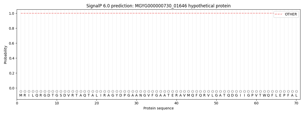
| Other | SP_Sec_SPI | LIPO_Sec_SPII | TAT_Tat_SPI | TATLIP_Sec_SPII | PILIN_Sec_SPIII |
|---|---|---|---|---|---|
| 0.998971 | 0.001041 | 0.000031 | 0.000003 | 0.000002 | 0.000004 |