You are browsing environment: HUMAN GUT
CAZyme Information: MGYG000000759_01039
You are here: Home > Sequence: MGYG000000759_01039
Basic Information |
Genomic context |
Full Sequence |
Enzyme annotations |
CAZy signature domains |
CDD domains |
CAZyme hits |
PDB hits |
Swiss-Prot hits |
SignalP and Lipop annotations |
TMHMM annotations
Basic Information help
| Species | ||||||||||||
|---|---|---|---|---|---|---|---|---|---|---|---|---|
| Lineage | Bacteria; Actinobacteriota; Actinomycetia; Mycobacteriales; Mycobacteriaceae; Corynebacterium; | |||||||||||
| CAZyme ID | MGYG000000759_01039 | |||||||||||
| CAZy Family | GT4 | |||||||||||
| CAZyme Description | Phosphatidyl-myo-inositol mannosyltransferase | |||||||||||
| CAZyme Property |
|
|||||||||||
| Genome Property |
|
|||||||||||
| Gene Location | Start: 954; End: 2057 Strand: + | |||||||||||
CDD Domains download full data without filtering help
| Cdd ID | Domain | E-Value | qStart | qEnd | sStart | sEnd | Domain Description |
|---|---|---|---|---|---|---|---|
| cd03801 | GT4_PimA-like | 2.85e-50 | 2 | 355 | 1 | 366 | phosphatidyl-myo-inositol mannosyltransferase. This family is most closely related to the GT4 family of glycosyltransferases and named after PimA in Propionibacterium freudenreichii, which is involved in the biosynthesis of phosphatidyl-myo-inositol mannosides (PIM) which are early precursors in the biosynthesis of lipomannans (LM) and lipoarabinomannans (LAM), and catalyzes the addition of a mannosyl residue from GDP-D-mannose (GDP-Man) to the position 2 of the carrier lipid phosphatidyl-myo-inositol (PI) to generate a phosphatidyl-myo-inositol bearing an alpha-1,2-linked mannose residue (PIM1). Glycosyltransferases catalyze the transfer of sugar moieties from activated donor molecules to specific acceptor molecules, forming glycosidic bonds. The acceptor molecule can be a lipid, a protein, a heterocyclic compound, or another carbohydrate residue. This group of glycosyltransferases is most closely related to the previously defined glycosyltransferase family 1 (GT1). The members of this family may transfer UDP, ADP, GDP, or CMP linked sugars. The diverse enzymatic activities among members of this family reflect a wide range of biological functions. The protein structure available for this family has the GTB topology, one of the two protein topologies observed for nucleotide-sugar-dependent glycosyltransferases. GTB proteins have distinct N- and C- terminal domains each containing a typical Rossmann fold. The two domains have high structural homology despite minimal sequence homology. The large cleft that separates the two domains includes the catalytic center and permits a high degree of flexibility. The members of this family are found mainly in certain bacteria and archaea. |
| COG0438 | RfaB | 4.91e-34 | 1 | 357 | 1 | 377 | Glycosyltransferase involved in cell wall bisynthesis [Cell wall/membrane/envelope biogenesis]. |
| cd03817 | GT4_UGDG-like | 9.43e-24 | 2 | 357 | 1 | 372 | UDP-Glc:1,2-diacylglycerol 3-a-glucosyltransferase and similar proteins. This family is most closely related to the GT1 family of glycosyltransferases. UDP-glucose-diacylglycerol glucosyltransferase (EC 2.4.1.337, UGDG; also known as 1,2-diacylglycerol 3-glucosyltransferase) catalyzes the transfer of glucose from UDP-glucose to 1,2-diacylglycerol forming 3-D-glucosyl-1,2-diacylglycerol. |
| cd03795 | GT4_WfcD-like | 1.20e-23 | 15 | 340 | 14 | 347 | Escherichia coli alpha-1,3-mannosyltransferase WfcD and similar proteins. This family is most closely related to the GT4 family of glycosyltransferases. Glycosyltransferases catalyze the transfer of sugar moieties from activated donor molecules to specific acceptor molecules, forming glycosidic bonds. The acceptor molecule can be a lipid, a protein, a heterocyclic compound, or another carbohydrate residue. This group of glycosyltransferases is most closely related to the previously defined glycosyltransferase family 1 (GT1). The members of this family may transfer UDP, ADP, GDP, or CMP-linked sugars. The diverse enzymatic activities among members of this family reflect a wide range of biological functions. The protein structure available for this family has the GTB topology, one of the two protein topologies observed for nucleotide-sugar-dependent glycosyltransferases. GTB proteins have distinct N- and C- terminal domains each containing a typical Rossmann fold. The two domains have high structural homology despite minimal sequence homology. The large cleft that separates the two domains includes the catalytic center and permits a high degree of flexibility. The members of this family are found mainly in bacteria and eukaryotes. |
| pfam13439 | Glyco_transf_4 | 6.11e-23 | 14 | 173 | 1 | 169 | Glycosyltransferase Family 4. |
CAZyme Hits help
| Hit ID | E-Value | Query Start | Query End | Hit Start | Hit End |
|---|---|---|---|---|---|
| QQN46738.1 | 9.06e-258 | 1 | 367 | 1 | 367 |
| QRQ66443.1 | 2.55e-248 | 1 | 367 | 1 | 367 |
| QQB00125.1 | 1.21e-246 | 1 | 367 | 1 | 367 |
| QQU81858.1 | 1.72e-246 | 1 | 367 | 1 | 367 |
| VEH73046.1 | 9.88e-230 | 1 | 367 | 1 | 367 |
PDB Hits download full data without filtering help
| Hit ID | E-Value | Query Start | Query End | Hit Start | Hit End | Description |
|---|---|---|---|---|---|---|
| 4N9W_A | 6.43e-118 | 1 | 365 | 5 | 376 | Crystalstructure of phosphatidyl mannosyltransferase PimA [Mycolicibacterium smegmatis MC2 155],4NC9_A Crystal structure of phosphatidyl mannosyltransferase PimA [Mycolicibacterium smegmatis MC2 155],4NC9_B Crystal structure of phosphatidyl mannosyltransferase PimA [Mycolicibacterium smegmatis MC2 155],4NC9_C Crystal structure of phosphatidyl mannosyltransferase PimA [Mycolicibacterium smegmatis MC2 155],4NC9_D Crystal structure of phosphatidyl mannosyltransferase PimA [Mycolicibacterium smegmatis MC2 155] |
| 2GEJ_A | 1.08e-117 | 1 | 365 | 21 | 392 | CrystalStructure of phosphatidylinositol mannosyltransferase (PimA) from Mycobacterium smegmatis in complex with GDP-Man [Mycolicibacterium smegmatis MC2 155],2GEK_A Crystal Structure of phosphatidylinositol mannosyltransferase (PimA) from Mycobacterium smegmatis in complex with GDP [Mycolicibacterium smegmatis MC2 155] |
| 6KIH_A | 1.14e-13 | 152 | 354 | 207 | 421 | Sucrose-phosphatesynthase (tll1590) from Thermosynechococcus elongatus [Thermosynechococcus vestitus],6KIH_B Sucrose-phosphate synthase (tll1590) from Thermosynechococcus elongatus [Thermosynechococcus vestitus],6KIH_C Sucrose-phosphate synthase (tll1590) from Thermosynechococcus elongatus [Thermosynechococcus vestitus],6KIH_D Sucrose-phosphate synthase (tll1590) from Thermosynechococcus elongatus [Thermosynechococcus vestitus],6KIH_E Sucrose-phosphate synthase (tll1590) from Thermosynechococcus elongatus [Thermosynechococcus vestitus],6KIH_F Sucrose-phosphate synthase (tll1590) from Thermosynechococcus elongatus [Thermosynechococcus vestitus],6KIH_G Sucrose-phosphate synthase (tll1590) from Thermosynechococcus elongatus [Thermosynechococcus vestitus],6KIH_H Sucrose-phosphate synthase (tll1590) from Thermosynechococcus elongatus [Thermosynechococcus vestitus],6KIH_I Sucrose-phosphate synthase (tll1590) from Thermosynechococcus elongatus [Thermosynechococcus vestitus],6KIH_J Sucrose-phosphate synthase (tll1590) from Thermosynechococcus elongatus [Thermosynechococcus vestitus],6KIH_K Sucrose-phosphate synthase (tll1590) from Thermosynechococcus elongatus [Thermosynechococcus vestitus],6KIH_L Sucrose-phosphate synthase (tll1590) from Thermosynechococcus elongatus [Thermosynechococcus vestitus] |
| 3C4Q_A | 2.60e-10 | 13 | 354 | 20 | 402 | Structureof the retaining glycosyltransferase MshA : The first step in mycothiol biosynthesis. Organism : Corynebacterium glutamicum- Complex with UDP [Corynebacterium glutamicum],3C4Q_B Structure of the retaining glycosyltransferase MshA : The first step in mycothiol biosynthesis. Organism : Corynebacterium glutamicum- Complex with UDP [Corynebacterium glutamicum],3C4V_A Structure of the retaining glycosyltransferase MshA:The first step in mycothiol biosynthesis. Organism: Corynebacterium glutamicum : Complex with UDP and 1L-INS-1-P. [Corynebacterium glutamicum],3C4V_B Structure of the retaining glycosyltransferase MshA:The first step in mycothiol biosynthesis. Organism: Corynebacterium glutamicum : Complex with UDP and 1L-INS-1-P. [Corynebacterium glutamicum] |
| 3C48_A | 2.67e-10 | 13 | 354 | 40 | 422 | Structureof the retaining glycosyltransferase MshA: The first step in mycothiol biosynthesis. Organism: Corynebacterium glutamicum- APO (OPEN) structure. [Corynebacterium glutamicum],3C48_B Structure of the retaining glycosyltransferase MshA: The first step in mycothiol biosynthesis. Organism: Corynebacterium glutamicum- APO (OPEN) structure. [Corynebacterium glutamicum] |
Swiss-Prot Hits download full data without filtering help
| Hit ID | E-Value | Query Start | Query End | Hit Start | Hit End | Description |
|---|---|---|---|---|---|---|
| A0QWG6 | 3.09e-117 | 1 | 365 | 1 | 372 | Phosphatidyl-myo-inositol mannosyltransferase OS=Mycolicibacterium smegmatis (strain ATCC 700084 / mc(2)155) OX=246196 GN=pimA PE=1 SV=1 |
| P9WMZ5 | 4.08e-113 | 1 | 365 | 1 | 376 | Phosphatidyl-myo-inositol mannosyltransferase OS=Mycobacterium tuberculosis (strain ATCC 25618 / H37Rv) OX=83332 GN=pimA PE=1 SV=1 |
| Q7TY88 | 4.08e-113 | 1 | 365 | 1 | 376 | Phosphatidyl-myo-inositol mannosyltransferase OS=Mycobacterium bovis (strain ATCC BAA-935 / AF2122/97) OX=233413 GN=pimA PE=3 SV=1 |
| P9WMZ4 | 4.08e-113 | 1 | 365 | 1 | 376 | Phosphatidyl-myo-inositol mannosyltransferase OS=Mycobacterium tuberculosis (strain CDC 1551 / Oshkosh) OX=83331 GN=pimA PE=3 SV=1 |
| O07147 | 1.07e-109 | 1 | 365 | 1 | 372 | Phosphatidyl-myo-inositol mannosyltransferase OS=Mycobacterium leprae (strain TN) OX=272631 GN=pimA PE=3 SV=1 |
SignalP and Lipop Annotations help
This protein is predicted as OTHER
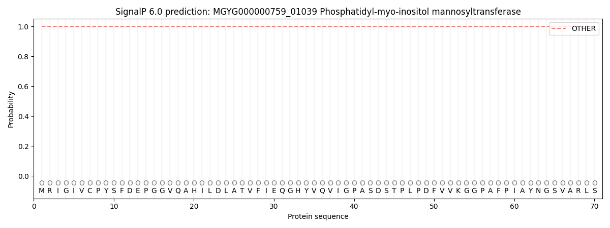
| Other | SP_Sec_SPI | LIPO_Sec_SPII | TAT_Tat_SPI | TATLIP_Sec_SPII | PILIN_Sec_SPIII |
|---|---|---|---|---|---|
| 1.000064 | 0.000000 | 0.000000 | 0.000000 | 0.000000 | 0.000000 |