You are browsing environment: HUMAN GUT
CAZyme Information: MGYG000000759_01390
You are here: Home > Sequence: MGYG000000759_01390
Basic Information |
Genomic context |
Full Sequence |
Enzyme annotations |
CAZy signature domains |
CDD domains |
CAZyme hits |
PDB hits |
Swiss-Prot hits |
SignalP and Lipop annotations |
TMHMM annotations
Basic Information help
| Species | ||||||||||||
|---|---|---|---|---|---|---|---|---|---|---|---|---|
| Lineage | Bacteria; Actinobacteriota; Actinomycetia; Mycobacteriales; Mycobacteriaceae; Corynebacterium; | |||||||||||
| CAZyme ID | MGYG000000759_01390 | |||||||||||
| CAZy Family | GH13 | |||||||||||
| CAZyme Description | Alpha-1,4-glucan:maltose-1-phosphate maltosyltransferase | |||||||||||
| CAZyme Property |
|
|||||||||||
| Genome Property |
|
|||||||||||
| Gene Location | Start: 902; End: 2917 Strand: - | |||||||||||
CAZyme Signature Domains help
| Family | Start | End | Evalue | family coverage |
|---|---|---|---|---|
| GH13 | 228 | 444 | 5.6e-84 | 0.9857142857142858 |
CDD Domains download full data without filtering help
| Cdd ID | Domain | E-Value | qStart | qEnd | sStart | sEnd | Domain Description |
|---|---|---|---|---|---|---|---|
| cd11344 | AmyAc_GlgE_like | 0.0 | 199 | 554 | 1 | 355 | Alpha amylase catalytic domain found in GlgE-like proteins. GlgE is a (1,4)-a-D-glucan:phosphate a-D-maltosyltransferase, involved in a-glucan biosynthesis in bacteria. It is also an anti-tuberculosis drug target. GlgE isoform I from Streptomyces coelicolor has the same catalytic and very similar kinetic properties to GlgE from Mycobacterium tuberculosis. GlgE from Streptomyces coelicolor forms a homodimer with each subunit comprising five domains (A, B, C, N, and S) and 2 inserts. Domain A is a catalytic alpha-amylase-type domain that along with domain N, which has a beta-sandwich fold and forms the core of the dimer interface, binds cyclodextrins. Domain A, B, and the 2 inserts define a well conserved donor pocket that binds maltose. Cyclodextrins competitively inhibit the binding of maltooligosaccharides to the S. coelicolor enzyme, indicating that the hydrophobic patch overlaps with the acceptor binding site. This is not the case in M. tuberculosis GlgE because cyclodextrins do not inhibit this enzyme, despite acceptor length specificity being conserved. Domain C is hypothesized to help stabilize domain A and could be involved in substrate binding. Domain S is a helix bundle that is inserted within the N domain and it plays a role in the dimer interface and interacts directly with domain B. The Alpha-amylase family comprises the largest family of glycoside hydrolases (GH), with the majority of enzymes acting on starch, glycogen, and related oligo- and polysaccharides. These proteins catalyze the transformation of alpha-1,4 and alpha-1,6 glucosidic linkages with retention of the anomeric center. The protein is described as having 3 domains: A, B, C. A is a (beta/alpha) 8-barrel; B is a loop between the beta 3 strand and alpha 3 helix of A; C is the C-terminal extension characterized by a Greek key. The majority of the enzymes have an active site cleft found between domains A and B where a triad of catalytic residues (Asp, Glu and Asp) performs catalysis. Other members of this family have lost the catalytic activity as in the case of the human 4F2hc, or only have 2 residues that serve as the catalytic nucleophile and the acid/base, such as Thermus A4 beta-galactosidase with 2 Glu residues (GH42) and human alpha-galactosidase with 2 Asp residues (GH31). The family members are quite extensive and include: alpha amylase, maltosyltransferase, cyclodextrin glycotransferase, maltogenic amylase, neopullulanase, isoamylase, 1,4-alpha-D-glucan maltotetrahydrolase, 4-alpha-glucotransferase, oligo-1,6-glucosidase, amylosucrase, sucrose phosphorylase, and amylomaltase. |
| pfam11896 | DUF3416 | 7.16e-65 | 6 | 194 | 2 | 185 | Domain of unknown function (DUF3416). This presumed domain is functionally uncharacterized. This domain is found in bacteria and archaea. This domain is about 190 amino acids in length. This domain is found associated with pfam00128. |
| cd11313 | AmyAc_arch_bac_AmyA | 7.06e-45 | 203 | 557 | 8 | 334 | Alpha amylase catalytic domain found in archaeal and bacterial Alpha-amylases (also called 1,4-alpha-D-glucan-4-glucanohydrolase). AmyA (EC 3.2.1.1) catalyzes the hydrolysis of alpha-(1,4) glycosidic linkages of glycogen, starch, related polysaccharides, and some oligosaccharides. This group includes firmicutes, bacteroidetes, and proteobacteria. The Alpha-amylase family comprises the largest family of glycoside hydrolases (GH), with the majority of enzymes acting on starch, glycogen, and related oligo- and polysaccharides. These proteins catalyze the transformation of alpha-1,4 and alpha-1,6 glucosidic linkages with retention of the anomeric center. The protein is described as having 3 domains: A, B, C. A is a (beta/alpha) 8-barrel; B is a loop between the beta 3 strand and alpha 3 helix of A; C is the C-terminal extension characterized by a Greek key. The majority of the enzymes have an active site cleft found between domains A and B where a triad of catalytic residues (Asp, Glu and Asp) performs catalysis. Other members of this family have lost the catalytic activity as in the case of the human 4F2hc, or only have 2 residues that serve as the catalytic nucleophile and the acid/base, such as Thermus A4 beta-galactosidase with 2 Glu residues (GH42) and human alpha-galactosidase with 2 Asp residues (GH31). The family members are quite extensive and include: alpha amylase, maltosyltransferase, cyclodextrin glycotransferase, maltogenic amylase, neopullulanase, isoamylase, 1,4-alpha-D-glucan maltotetrahydrolase, 4-alpha-glucotransferase, oligo-1,6-glucosidase, amylosucrase, sucrose phosphorylase, and amylomaltase. |
| COG0366 | AmyA | 4.18e-20 | 202 | 454 | 3 | 277 | Glycosidase [Carbohydrate transport and metabolism]. |
| cd00551 | AmyAc_family | 1.52e-19 | 203 | 467 | 3 | 215 | Alpha amylase catalytic domain family. The Alpha-amylase family comprises the largest family of glycoside hydrolases (GH), with the majority of enzymes acting on starch, glycogen, and related oligo- and polysaccharides. These proteins catalyze the transformation of alpha-1,4 and alpha-1,6 glucosidic linkages with retention of the anomeric center. The protein is described as having 3 domains: A, B, C. A is a (beta/alpha) 8-barrel; B is a loop between the beta 3 strand and alpha 3 helix of A; and C is the C-terminal extension characterized by a Greek key. The majority of the enzymes have an active site cleft found between domains A and B where a triad of catalytic residues (Asp, Glu and Asp) performs catalysis. Other members of this family have lost this catalytic activity as in the case of the human 4F2hc, or only have 2 residues that serve as the catalytic nucleophile and the acid/base, such as Thermus A4 beta-galactosidase with 2 Glu residues (GH42) and human alpha-galactosidase with 2 Asp residues (GH31). The family members are quite extensive and include: alpha amylase, maltosyltransferase, cyclodextrin glycotransferase, maltogenic amylase, neopullulanase, isoamylase, 1,4-alpha-D-glucan maltotetrahydrolase, 4-alpha-glucotransferase, oligo-1,6-glucosidase, amylosucrase, sucrose phosphorylase, and amylomaltase. |
CAZyme Hits help
| Hit ID | E-Value | Query Start | Query End | Hit Start | Hit End |
|---|---|---|---|---|---|
| QQN47035.1 | 0.0 | 3 | 671 | 1 | 669 |
| QQU81566.1 | 0.0 | 3 | 671 | 1 | 669 |
| QRQ66738.1 | 0.0 | 3 | 671 | 1 | 669 |
| QQA99823.1 | 0.0 | 3 | 671 | 1 | 669 |
| QQU95099.1 | 0.0 | 3 | 671 | 1 | 668 |
PDB Hits download full data without filtering help
| Hit ID | E-Value | Query Start | Query End | Hit Start | Hit End | Description |
|---|---|---|---|---|---|---|
| 5CGM_A | 4.49e-281 | 6 | 658 | 6 | 693 | Structureof Mycobacterium thermoresistibile GlgE in complex with maltose at 1.95A resolution [Mycolicibacterium thermoresistibile ATCC 19527],5CGM_B Structure of Mycobacterium thermoresistibile GlgE in complex with maltose at 1.95A resolution [Mycolicibacterium thermoresistibile ATCC 19527],5CIM_A Structure of Mycobacterium thermoresistibile GlgE in complex with maltose (cocrystallisation with maltose-1-phosphate) at 3.32A resolution [Mycolicibacterium thermoresistibile ATCC 19527],5CIM_B Structure of Mycobacterium thermoresistibile GlgE in complex with maltose (cocrystallisation with maltose-1-phosphate) at 3.32A resolution [Mycolicibacterium thermoresistibile ATCC 19527],5CJ5_A Structure of Mycobacterium thermoresistibile GlgE APO form at 3.13A resolution [Mycolicibacterium thermoresistibile ATCC 19527],5CJ5_B Structure of Mycobacterium thermoresistibile GlgE APO form at 3.13A resolution [Mycolicibacterium thermoresistibile ATCC 19527] |
| 4U33_A | 1.69e-267 | 6 | 664 | 39 | 722 | Structureof Mtb GlgE bound to maltose [Mycobacterium tuberculosis CDC1551],4U33_B Structure of Mtb GlgE bound to maltose [Mycobacterium tuberculosis CDC1551],4U33_C Structure of Mtb GlgE bound to maltose [Mycobacterium tuberculosis CDC1551],4U33_D Structure of Mtb GlgE bound to maltose [Mycobacterium tuberculosis CDC1551],4U33_E Structure of Mtb GlgE bound to maltose [Mycobacterium tuberculosis CDC1551],4U33_F Structure of Mtb GlgE bound to maltose [Mycobacterium tuberculosis CDC1551],4U3C_A Docking Site of Maltohexaose in the Mtb GlgE [Mycobacterium tuberculosis CDC1551],4U3C_B Docking Site of Maltohexaose in the Mtb GlgE [Mycobacterium tuberculosis CDC1551],4U3C_C Docking Site of Maltohexaose in the Mtb GlgE [Mycobacterium tuberculosis CDC1551],4U3C_D Docking Site of Maltohexaose in the Mtb GlgE [Mycobacterium tuberculosis CDC1551],4U3C_E Docking Site of Maltohexaose in the Mtb GlgE [Mycobacterium tuberculosis CDC1551],4U3C_F Docking Site of Maltohexaose in the Mtb GlgE [Mycobacterium tuberculosis CDC1551] |
| 5VSJ_A | 4.85e-219 | 6 | 647 | 20 | 658 | ScoGlgEI-V279S in complex with a pyrolidene-based ethyl-phosphonate compound [Streptomyces coelicolor A3(2)],5VSJ_B Sco GlgEI-V279S in complex with a pyrolidene-based ethyl-phosphonate compound [Streptomyces coelicolor A3(2)],5VT4_A Sco GlgEI-V279S in complex with a pyrolidene-based methyl-phosphonate compound [Streptomyces coelicolor A3(2)],5VT4_B Sco GlgEI-V279S in complex with a pyrolidene-based methyl-phosphonate compound [Streptomyces coelicolor A3(2)],5VT4_C Sco GlgEI-V279S in complex with a pyrolidene-based methyl-phosphonate compound [Streptomyces coelicolor A3(2)],5VT4_D Sco GlgEI-V279S in complex with a pyrolidene-based methyl-phosphonate compound [Streptomyces coelicolor A3(2)] |
| 4U2Y_A | 6.73e-219 | 6 | 655 | 20 | 666 | ScoGlgEI-V279S in Complex with Reaction Intermediate Azasugar [Streptomyces coelicolor A3(2)],4U2Y_B Sco GlgEI-V279S in Complex with Reaction Intermediate Azasugar [Streptomyces coelicolor A3(2)],4U2Z_A X-ray crystal structure of an Sco GlgEI-V279S/1,2,2-trifluromaltose complex [Streptomyces coelicolor A3(2)],4U2Z_B X-ray crystal structure of an Sco GlgEI-V279S/1,2,2-trifluromaltose complex [Streptomyces coelicolor A3(2)],4U31_A Sco GlgEI-V279S in Complex with maltose-C-phosphonate [Streptomyces coelicolor A3(2)],4U31_B Sco GlgEI-V279S in Complex with maltose-C-phosphonate [Streptomyces coelicolor A3(2)],7MEL_A Chain A, Alpha-1,4-glucan:maltose-1-phosphate maltosyltransferase 1 [Streptomyces coelicolor A3(2)],7MEL_B Chain B, Alpha-1,4-glucan:maltose-1-phosphate maltosyltransferase 1 [Streptomyces coelicolor A3(2)],7MGY_A Chain A, Alpha-1,4-glucan:maltose-1-phosphate maltosyltransferase 1 [Streptomyces coelicolor A3(2)],7MGY_B Chain B, Alpha-1,4-glucan:maltose-1-phosphate maltosyltransferase 1 [Streptomyces coelicolor A3(2)] |
| 3ZSS_A | 8.13e-218 | 6 | 655 | 40 | 686 | Apoform of GlgE isoform 1 from Streptomyces coelicolor [Streptomyces coelicolor],3ZSS_B Apo form of GlgE isoform 1 from Streptomyces coelicolor [Streptomyces coelicolor],3ZSS_C Apo form of GlgE isoform 1 from Streptomyces coelicolor [Streptomyces coelicolor],3ZSS_D Apo form of GlgE isoform 1 from Streptomyces coelicolor [Streptomyces coelicolor],3ZST_A GlgE isoform 1 from Streptomyces coelicolor with alpha-cyclodextrin bound [Streptomyces coelicolor],3ZST_B GlgE isoform 1 from Streptomyces coelicolor with alpha-cyclodextrin bound [Streptomyces coelicolor],3ZT5_A GlgE isoform 1 from Streptomyces coelicolor with maltose bound [Streptomyces coelicolor],3ZT5_B GlgE isoform 1 from Streptomyces coelicolor with maltose bound [Streptomyces coelicolor],3ZT5_C GlgE isoform 1 from Streptomyces coelicolor with maltose bound [Streptomyces coelicolor],3ZT5_D GlgE isoform 1 from Streptomyces coelicolor with maltose bound [Streptomyces coelicolor],3ZT6_A GlgE isoform 1 from Streptomyces coelicolor with alpha-cyclodextrin and maltose bound [Streptomyces coelicolor],3ZT6_B GlgE isoform 1 from Streptomyces coelicolor with alpha-cyclodextrin and maltose bound [Streptomyces coelicolor],3ZT6_C GlgE isoform 1 from Streptomyces coelicolor with alpha-cyclodextrin and maltose bound [Streptomyces coelicolor],3ZT6_D GlgE isoform 1 from Streptomyces coelicolor with alpha-cyclodextrin and maltose bound [Streptomyces coelicolor],3ZT7_A GlgE isoform 1 from Streptomyces coelicolor with beta-cyclodextrin and maltose bound [Streptomyces coelicolor],3ZT7_B GlgE isoform 1 from Streptomyces coelicolor with beta-cyclodextrin and maltose bound [Streptomyces coelicolor],3ZT7_C GlgE isoform 1 from Streptomyces coelicolor with beta-cyclodextrin and maltose bound [Streptomyces coelicolor],3ZT7_D GlgE isoform 1 from Streptomyces coelicolor with beta-cyclodextrin and maltose bound [Streptomyces coelicolor] |
Swiss-Prot Hits download full data without filtering help
| Hit ID | E-Value | Query Start | Query End | Hit Start | Hit End | Description |
|---|---|---|---|---|---|---|
| Q8NR39 | 3.68e-315 | 3 | 670 | 1 | 674 | Alpha-1,4-glucan:maltose-1-phosphate maltosyltransferase OS=Corynebacterium glutamicum (strain ATCC 13032 / DSM 20300 / BCRC 11384 / JCM 1318 / LMG 3730 / NCIMB 10025) OX=196627 GN=glgE PE=3 SV=2 |
| D9Q7U8 | 3.28e-310 | 3 | 669 | 1 | 669 | Alpha-1,4-glucan:maltose-1-phosphate maltosyltransferase OS=Corynebacterium pseudotuberculosis (strain 1002) OX=679896 GN=glgE PE=3 SV=2 |
| Q9RP48 | 1.78e-282 | 6 | 665 | 9 | 697 | Alpha-1,4-glucan:maltose-1-phosphate maltosyltransferase OS=Mycolicibacterium smegmatis (strain ATCC 700084 / mc(2)155) OX=246196 GN=glgE PE=1 SV=1 |
| P9WQ17 | 4.28e-267 | 6 | 664 | 17 | 700 | Alpha-1,4-glucan:maltose-1-phosphate maltosyltransferase OS=Mycobacterium tuberculosis (strain ATCC 25618 / H37Rv) OX=83332 GN=glgE PE=1 SV=1 |
| P9WQ16 | 4.28e-267 | 6 | 664 | 17 | 700 | Alpha-1,4-glucan:maltose-1-phosphate maltosyltransferase OS=Mycobacterium tuberculosis (strain CDC 1551 / Oshkosh) OX=83331 GN=glgE PE=1 SV=1 |
SignalP and Lipop Annotations help
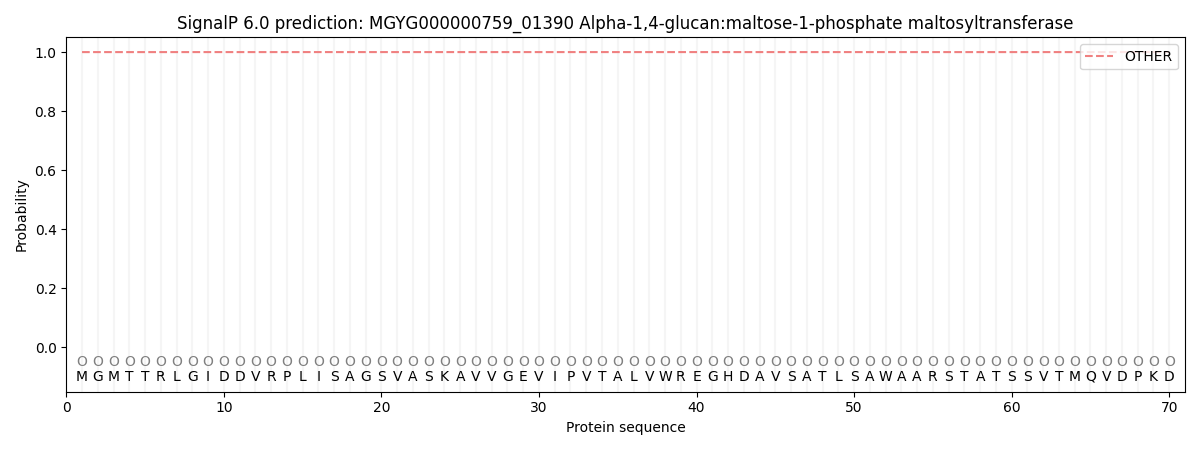
This protein is predicted as OTHER
| Other | SP_Sec_SPI | LIPO_Sec_SPII | TAT_Tat_SPI | TATLIP_Sec_SPII | PILIN_Sec_SPIII |
|---|---|---|---|---|---|
| 1.000045 | 0.000004 | 0.000000 | 0.000000 | 0.000000 | 0.000000 |