You are browsing environment: HUMAN GUT
CAZyme Information: MGYG000000792_00108
You are here: Home > Sequence: MGYG000000792_00108
Basic Information |
Genomic context |
Full Sequence |
Enzyme annotations |
CAZy signature domains |
CDD domains |
CAZyme hits |
PDB hits |
Swiss-Prot hits |
SignalP and Lipop annotations |
TMHMM annotations
Basic Information help
| Species | Eubacterium_M sp900540015 | |||||||||||
|---|---|---|---|---|---|---|---|---|---|---|---|---|
| Lineage | Bacteria; Firmicutes_A; Clostridia; Peptostreptococcales; Anaerovoracaceae; Eubacterium_M; Eubacterium_M sp900540015 | |||||||||||
| CAZyme ID | MGYG000000792_00108 | |||||||||||
| CAZy Family | GT4 | |||||||||||
| CAZyme Description | Ubiquinone biosynthesis O-methyltransferase | |||||||||||
| CAZyme Property |
|
|||||||||||
| Genome Property |
|
|||||||||||
| Gene Location | Start: 120133; End: 125058 Strand: + | |||||||||||
CDD Domains download full data without filtering help
| Cdd ID | Domain | E-Value | qStart | qEnd | sStart | sEnd | Domain Description |
|---|---|---|---|---|---|---|---|
| cd03801 | GT4_PimA-like | 1.74e-21 | 1291 | 1631 | 31 | 366 | phosphatidyl-myo-inositol mannosyltransferase. This family is most closely related to the GT4 family of glycosyltransferases and named after PimA in Propionibacterium freudenreichii, which is involved in the biosynthesis of phosphatidyl-myo-inositol mannosides (PIM) which are early precursors in the biosynthesis of lipomannans (LM) and lipoarabinomannans (LAM), and catalyzes the addition of a mannosyl residue from GDP-D-mannose (GDP-Man) to the position 2 of the carrier lipid phosphatidyl-myo-inositol (PI) to generate a phosphatidyl-myo-inositol bearing an alpha-1,2-linked mannose residue (PIM1). Glycosyltransferases catalyze the transfer of sugar moieties from activated donor molecules to specific acceptor molecules, forming glycosidic bonds. The acceptor molecule can be a lipid, a protein, a heterocyclic compound, or another carbohydrate residue. This group of glycosyltransferases is most closely related to the previously defined glycosyltransferase family 1 (GT1). The members of this family may transfer UDP, ADP, GDP, or CMP linked sugars. The diverse enzymatic activities among members of this family reflect a wide range of biological functions. The protein structure available for this family has the GTB topology, one of the two protein topologies observed for nucleotide-sugar-dependent glycosyltransferases. GTB proteins have distinct N- and C- terminal domains each containing a typical Rossmann fold. The two domains have high structural homology despite minimal sequence homology. The large cleft that separates the two domains includes the catalytic center and permits a high degree of flexibility. The members of this family are found mainly in certain bacteria and archaea. |
| COG1196 | Smc | 2.43e-18 | 236 | 477 | 667 | 904 | Chromosome segregation ATPase [Cell cycle control, cell division, chromosome partitioning]. |
| TIGR02168 | SMC_prok_B | 1.19e-17 | 212 | 506 | 236 | 517 | chromosome segregation protein SMC, common bacterial type. SMC (structural maintenance of chromosomes) proteins bind DNA and act in organizing and segregating chromosomes for partition. SMC proteins are found in bacteria, archaea, and eukaryotes. This family represents the SMC protein of most bacteria. The smc gene is often associated with scpB (TIGR00281) and scpA genes, where scp stands for segregation and condensation protein. SMC was shown (in Caulobacter crescentus) to be induced early in S phase but present and bound to DNA throughout the cell cycle. [Cellular processes, Cell division, DNA metabolism, Chromosome-associated proteins] |
| COG1196 | Smc | 3.78e-16 | 224 | 491 | 234 | 515 | Chromosome segregation ATPase [Cell cycle control, cell division, chromosome partitioning]. |
| TIGR02168 | SMC_prok_B | 3.03e-15 | 238 | 520 | 195 | 499 | chromosome segregation protein SMC, common bacterial type. SMC (structural maintenance of chromosomes) proteins bind DNA and act in organizing and segregating chromosomes for partition. SMC proteins are found in bacteria, archaea, and eukaryotes. This family represents the SMC protein of most bacteria. The smc gene is often associated with scpB (TIGR00281) and scpA genes, where scp stands for segregation and condensation protein. SMC was shown (in Caulobacter crescentus) to be induced early in S phase but present and bound to DNA throughout the cell cycle. [Cellular processes, Cell division, DNA metabolism, Chromosome-associated proteins] |
CAZyme Hits help
| Hit ID | E-Value | Query Start | Query End | Hit Start | Hit End |
|---|---|---|---|---|---|
| AIY81850.1 | 0.0 | 573 | 1630 | 32 | 1099 |
| SDS87966.1 | 1.21e-281 | 7 | 1629 | 25 | 1608 |
| AZN37304.1 | 3.32e-273 | 7 | 1630 | 25 | 1888 |
| ALS25943.1 | 8.86e-273 | 595 | 1633 | 50 | 1100 |
| QXH87868.1 | 3.47e-269 | 7 | 1630 | 21 | 1822 |
PDB Hits download full data without filtering help
| Hit ID | E-Value | Query Start | Query End | Hit Start | Hit End | Description |
|---|---|---|---|---|---|---|
| 3MGG_A | 2.50e-06 | 38 | 138 | 38 | 140 | CrystalStructure of Methyl Transferase from Methanosarcina mazei [Methanosarcina mazei],3MGG_B Crystal Structure of Methyl Transferase from Methanosarcina mazei [Methanosarcina mazei] |
| 6GKV_A | 2.88e-06 | 38 | 138 | 124 | 225 | Crystalstructure of Coclaurine N-Methyltransferase (CNMT) bound to N-methylheliamine and SAH [Coptis japonica],6GKV_B Crystal structure of Coclaurine N-Methyltransferase (CNMT) bound to N-methylheliamine and SAH [Coptis japonica],6GKY_A Crystal structure of Coclaurine N-Methyltransferase (CNMT) bound to N-methylheliamine and SAH [Coptis japonica],6GKY_B Crystal structure of Coclaurine N-Methyltransferase (CNMT) bound to N-methylheliamine and SAH [Coptis japonica] |
| 6GKZ_A | 6.68e-06 | 38 | 138 | 124 | 225 | Crystalstructure of Coclaurine N-Methyltransferase (CNMT) bound to N-methylheliamine and SAH [Coptis japonica],6GKZ_B Crystal structure of Coclaurine N-Methyltransferase (CNMT) bound to N-methylheliamine and SAH [Coptis japonica],6GKZ_C Crystal structure of Coclaurine N-Methyltransferase (CNMT) bound to N-methylheliamine and SAH [Coptis japonica],6GKZ_D Crystal structure of Coclaurine N-Methyltransferase (CNMT) bound to N-methylheliamine and SAH [Coptis japonica] |
Swiss-Prot Hits download full data without filtering help
| Hit ID | E-Value | Query Start | Query End | Hit Start | Hit End | Description |
|---|---|---|---|---|---|---|
| O31681 | 3.73e-07 | 630 | 932 | 42 | 341 | Spore protein YkvP OS=Bacillus subtilis (strain 168) OX=224308 GN=ykvP PE=2 SV=1 |
| A8HVC4 | 2.60e-06 | 38 | 139 | 65 | 165 | Ubiquinone biosynthesis O-methyltransferase OS=Azorhizobium caulinodans (strain ATCC 43989 / DSM 5975 / JCM 20966 / LMG 6465 / NBRC 14845 / NCIMB 13405 / ORS 571) OX=438753 GN=ubiG PE=3 SV=1 |
| Q108P1 | 5.26e-06 | 38 | 174 | 129 | 256 | (S)-tetrahydroprotoberberine N-methyltransferase OS=Papaver somniferum OX=3469 PE=1 SV=1 |
| Q5C9L6 | 5.33e-06 | 38 | 138 | 132 | 233 | (S)-coclaurine N-methyltransferase OS=Thalictrum flavum subsp. glaucum OX=150095 PE=1 SV=1 |
| Q9PAM5 | 5.82e-06 | 28 | 139 | 44 | 157 | Ubiquinone biosynthesis O-methyltransferase OS=Xylella fastidiosa (strain 9a5c) OX=160492 GN=ubiG PE=3 SV=1 |
SignalP and Lipop Annotations help
This protein is predicted as OTHER
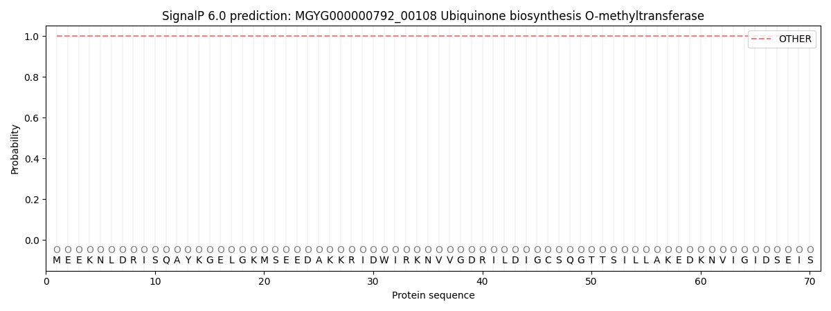
| Other | SP_Sec_SPI | LIPO_Sec_SPII | TAT_Tat_SPI | TATLIP_Sec_SPII | PILIN_Sec_SPIII |
|---|---|---|---|---|---|
| 1.000052 | 0.000000 | 0.000000 | 0.000000 | 0.000000 | 0.000000 |