You are browsing environment: HUMAN GUT
CAZyme Information: MGYG000000864_00205
You are here: Home > Sequence: MGYG000000864_00205
Basic Information |
Genomic context |
Full Sequence |
Enzyme annotations |
CAZy signature domains |
CDD domains |
CAZyme hits |
PDB hits |
Swiss-Prot hits |
SignalP and Lipop annotations |
TMHMM annotations
Basic Information help
| Species | CAG-632 sp900539185 | |||||||||||
|---|---|---|---|---|---|---|---|---|---|---|---|---|
| Lineage | Bacteria; Firmicutes_A; Clostridia; Lachnospirales; Lachnospiraceae; CAG-632; CAG-632 sp900539185 | |||||||||||
| CAZyme ID | MGYG000000864_00205 | |||||||||||
| CAZy Family | GH25 | |||||||||||
| CAZyme Description | hypothetical protein | |||||||||||
| CAZyme Property |
|
|||||||||||
| Genome Property |
|
|||||||||||
| Gene Location | Start: 212990; End: 214720 Strand: - | |||||||||||
CAZyme Signature Domains help
| Family | Start | End | Evalue | family coverage |
|---|---|---|---|---|
| GH25 | 193 | 379 | 4.5e-45 | 0.9943502824858758 |
CDD Domains download full data without filtering help
| Cdd ID | Domain | E-Value | qStart | qEnd | sStart | sEnd | Domain Description |
|---|---|---|---|---|---|---|---|
| cd06414 | GH25_LytC-like | 6.79e-80 | 192 | 389 | 3 | 191 | The LytC lysozyme of Streptococcus pneumoniae is a bacterial cell wall hydrolase that cleaves the beta1-4-glycosydic bond located between the N-acetylmuramoyl-N-glucosaminyl residues of the cell wall polysaccharide chains. LytC is composed of a C-terminal glycosyl hydrolase family 25 (GH25) domain and an N-terminal choline-binding module (CBM) consisting of eleven homologous repeats that specifically recognizes the choline residues of pneumococcal lipoteichoic and teichoic acids. This domain arrangement is the reverse of the major pneumococcal autolysin, LytA, and the CPL-1-like lytic enzymes of the pneumococcal bacteriophages, in which the CBM (consisting of six repeats) is at the C-terminus. This model represents the C-terminal catalytic domain of the LytC-like enzymes. |
| cd00599 | GH25_muramidase | 1.85e-37 | 192 | 387 | 2 | 185 | Endo-N-acetylmuramidases (muramidases) are lysozymes (also referred to as peptidoglycan hydrolases) that degrade bacterial cell walls by catalyzing the hydrolysis of 1,4-beta-linkages between N-acetylmuramic acid and N-acetyl-D-glucosamine residues. This family of muramidases contains a glycosyl hydrolase family 25 (GH25) catalytic domain and is found in bacteria, fungi, slime molds, round worms, protozoans and bacteriophages. The bacteriophage members are referred to as endolysins which are involved in lysing the host cell at the end of the replication cycle to allow release of mature phage particles. Endolysins are typically modular enzymes consisting of a catalytically active domain that hydrolyzes the peptidoglycan cell wall and a cell wall-binding domain that anchors the protein to the cell wall. Endolysins generally have narrow substrate specificities with either intra-species or intra-genus bacteriolytic activity. |
| NF033483 | PknB_PASTA_kin | 3.23e-35 | 400 | 535 | 427 | 563 | Stk1 family PASTA domain-containing Ser/Thr kinase. |
| NF033483 | PknB_PASTA_kin | 6.27e-35 | 398 | 539 | 356 | 499 | Stk1 family PASTA domain-containing Ser/Thr kinase. |
| pfam01183 | Glyco_hydro_25 | 3.54e-32 | 193 | 379 | 1 | 180 | Glycosyl hydrolases family 25. |
CAZyme Hits help
| Hit ID | E-Value | Query Start | Query End | Hit Start | Hit End |
|---|---|---|---|---|---|
| ADL33290.1 | 4.70e-53 | 150 | 389 | 989 | 1225 |
| BCK81546.1 | 4.78e-53 | 150 | 389 | 226 | 472 |
| AVM48757.1 | 2.42e-49 | 174 | 389 | 4 | 214 |
| QHI72152.1 | 6.24e-48 | 192 | 392 | 95 | 291 |
| ADK67502.1 | 1.91e-47 | 173 | 387 | 169 | 378 |
PDB Hits download full data without filtering help
| Hit ID | E-Value | Query Start | Query End | Hit Start | Hit End | Description |
|---|---|---|---|---|---|---|
| 2WW5_A | 2.29e-17 | 175 | 389 | 250 | 468 | 3D-structureof the modular autolysin LytC from Streptococcus pneumoniae at 1.6 A resolution [Streptococcus pneumoniae R6],2WWD_A 3D-structure of the modular autolysin LytC from Streptococcus pneumoniae in complex with pneummococcal peptidoglycan fragment [Streptococcus pneumoniae R6] |
| 2WWC_A | 5.41e-17 | 175 | 389 | 250 | 468 | 3D-structureof the modular autolysin LytC from Streptococcus pneumoniae in complex with synthetic peptidoglycan ligand [Streptococcus pneumoniae R6] |
| 1JFX_A | 1.47e-16 | 192 | 386 | 7 | 200 | Crystalstructure of the bacterial lysozyme from Streptomyces coelicolor at 1.65 A resolution [Streptomyces coelicolor] |
| 4KRU_A | 7.95e-14 | 192 | 386 | 22 | 205 | X-raystructure of catalytic domain of endolysin from clostridium perfringens phage phiSM101 [Clostridium phage phiSM101] |
| 4KRT_A | 3.54e-13 | 192 | 386 | 22 | 205 | X-raystructure of endolysin from clostridium perfringens phage phiSM101 [Clostridium phage phiSM101],4KRT_B X-ray structure of endolysin from clostridium perfringens phage phiSM101 [Clostridium phage phiSM101] |
Swiss-Prot Hits download full data without filtering help
| Hit ID | E-Value | Query Start | Query End | Hit Start | Hit End | Description |
|---|---|---|---|---|---|---|
| P25310 | 2.91e-15 | 192 | 386 | 84 | 277 | Lysozyme M1 OS=Streptomyces globisporus OX=1908 GN=acm PE=1 SV=1 |
| Q8FUI5 | 5.03e-12 | 398 | 538 | 372 | 514 | Probable serine/threonine-protein kinase CE0033 OS=Corynebacterium efficiens (strain DSM 44549 / YS-314 / AJ 12310 / JCM 11189 / NBRC 100395) OX=196164 GN=CE0033 PE=3 SV=1 |
| Q8R9T6 | 5.46e-10 | 409 | 535 | 417 | 544 | Probable serine/threonine-protein kinase Sps1 OS=Caldanaerobacter subterraneus subsp. tengcongensis (strain DSM 15242 / JCM 11007 / NBRC 100824 / MB4) OX=273068 GN=sps1 PE=3 SV=1 |
| Q8G4G1 | 8.06e-10 | 409 | 498 | 621 | 711 | Probable serine/threonine-protein kinase pknA2 OS=Bifidobacterium longum (strain NCC 2705) OX=206672 GN=pknA2 PE=3 SV=1 |
| Q9KIG4 | 9.67e-10 | 406 | 538 | 445 | 578 | Serine/threonine-protein kinase PK-1 OS=Streptomyces toyocaensis OX=55952 GN=spk1 PE=1 SV=1 |
SignalP and Lipop Annotations help
This protein is predicted as OTHER
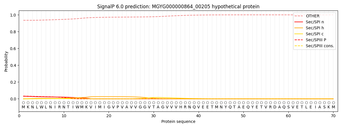
| Other | SP_Sec_SPI | LIPO_Sec_SPII | TAT_Tat_SPI | TATLIP_Sec_SPII | PILIN_Sec_SPIII |
|---|---|---|---|---|---|
| 0.936462 | 0.028160 | 0.001930 | 0.000225 | 0.000136 | 0.033121 |
