You are browsing environment: HUMAN GUT
CAZyme Information: MGYG000000971_00651
You are here: Home > Sequence: MGYG000000971_00651
Basic Information |
Genomic context |
Full Sequence |
Enzyme annotations |
CAZy signature domains |
CDD domains |
CAZyme hits |
PDB hits |
Swiss-Prot hits |
SignalP and Lipop annotations |
TMHMM annotations
Basic Information help
| Species | UMGS1518 sp900552575 | |||||||||||
|---|---|---|---|---|---|---|---|---|---|---|---|---|
| Lineage | Bacteria; Verrucomicrobiota; Lentisphaeria; Victivallales; Victivallaceae; UMGS1518; UMGS1518 sp900552575 | |||||||||||
| CAZyme ID | MGYG000000971_00651 | |||||||||||
| CAZy Family | GH20 | |||||||||||
| CAZyme Description | hypothetical protein | |||||||||||
| CAZyme Property |
|
|||||||||||
| Genome Property |
|
|||||||||||
| Gene Location | Start: 8910; End: 10763 Strand: - | |||||||||||
CAZyme Signature Domains help
| Family | Start | End | Evalue | family coverage |
|---|---|---|---|---|
| GH20 | 126 | 441 | 2.7e-40 | 0.9673590504451038 |
CDD Domains download full data without filtering help
| Cdd ID | Domain | E-Value | qStart | qEnd | sStart | sEnd | Domain Description |
|---|---|---|---|---|---|---|---|
| cd06565 | GH20_GcnA-like | 3.96e-84 | 132 | 441 | 1 | 301 | Glycosyl hydrolase family 20 (GH20) catalytic domain of N-acetyl-beta-D-glucosaminidase (GcnA, also known as BhsA) and related proteins. GcnA is an exoglucosidase which cleaves N-acetyl-beta-D-galactosamine (NAG) and N-acetyl-beta-D-galactosamine residues from 4-methylumbelliferylated (4MU) substrates, as well as cleaving NAG from chito-oligosaccharides (i.e. NAG polymers). In contrast, sulfated forms of the substrate are unable to be cleaved and act instead as mild competitive inhibitors. Additionally, the enzyme is known to be poisoned by several first-row transition metals as well as by mercury. GcnA forms a homodimer with subunits comprised of three domains, an N-terminal zincin-like domain, this central catalytic GH20 domain, and a C-terminal alpha helical domain. The GH20 hexosaminidases are thought to act via a catalytic mechanism in which the catalytic nucleophile is not provided by solvent or the enzyme, but by the substrate itself. |
| cd02742 | GH20_hexosaminidase | 1.11e-18 | 133 | 355 | 2 | 212 | Beta-N-acetylhexosaminidases of glycosyl hydrolase family 20 (GH20) catalyze the removal of beta-1,4-linked N-acetyl-D-hexosamine residues from the non-reducing ends of N-acetyl-beta-D-hexosaminides including N-acetylglucosides and N-acetylgalactosides. These enzymes are broadly distributed in microorganisms, plants and animals, and play roles in various key physiological and pathological processes. These processes include cell structural integrity, energy storage, cellular signaling, fertilization, pathogen defense, viral penetration, the development of carcinomas, inflammatory events and lysosomal storage disorders. The GH20 enzymes include the eukaryotic beta-N-acetylhexosaminidases A and B, the bacterial chitobiases, dispersin B, and lacto-N-biosidase. The GH20 hexosaminidases are thought to act via a catalytic mechanism in which the catalytic nucleophile is not provided by the solvent or the enzyme, but by the substrate itself. |
| cd06564 | GH20_DspB_LnbB-like | 7.55e-15 | 131 | 355 | 1 | 224 | Glycosyl hydrolase family 20 (GH20) catalytic domain of dispersin B (DspB), lacto-N-biosidase (LnbB) and related proteins. Dispersin B is a soluble beta-N-acetylglucosamidase found in bacteria that hydrolyzes the beta-1,6-linkages of PGA (poly-beta-(1,6)-N-acetylglucosamine), a major component of the extracellular polysaccharide matrix. Lacto-N-biosidase hydrolyzes lacto-N-biose (LNB) type I oligosaccharides at the nonreducing terminus to produce lacto-N-biose as part of the GNB/LNB (galacto-N-biose/lacto-N-biose I) degradation pathway. The lacto-N-biosidase from Bifidobacterium bifidum has this GH20 domain, a carbohydrate binding module 32, and a bacterial immunoglobulin-like domain 2, as well as a YSIRK signal peptide and a G5 membrane anchor at the N and C termini, respectively. The GH20 hexosaminidases are thought to act via a catalytic mechanism in which the catalytic nucleophile is not provided by solvent or the enzyme, but by the substrate itself. |
| COG3525 | Chb | 1.16e-12 | 41 | 334 | 173 | 491 | N-acetyl-beta-hexosaminidase [Carbohydrate transport and metabolism]. |
| pfam02838 | Glyco_hydro_20b | 6.06e-12 | 3 | 126 | 4 | 123 | Glycosyl hydrolase family 20, domain 2. This domain has a zincin-like fold. |
CAZyme Hits help
| Hit ID | E-Value | Query Start | Query End | Hit Start | Hit End |
|---|---|---|---|---|---|
| AUS98665.1 | 4.54e-140 | 80 | 605 | 77 | 612 |
| APC41628.1 | 2.48e-133 | 1 | 485 | 1 | 482 |
| QAS62105.1 | 8.73e-133 | 67 | 560 | 68 | 556 |
| AYE33954.1 | 8.73e-133 | 67 | 560 | 68 | 556 |
| ATD58120.1 | 3.46e-132 | 67 | 599 | 68 | 603 |
PDB Hits download full data without filtering help
| Hit ID | E-Value | Query Start | Query End | Hit Start | Hit End | Description |
|---|---|---|---|---|---|---|
| 6JQF_A | 2.15e-13 | 2 | 377 | 90 | 471 | Crystallizationanalysis of a beta-N-acetylhexosaminidase (Am2136) from Akkermansia muciniphila [Akkermansia muciniphila ATCC BAA-835] |
| 2GK1_I | 1.46e-11 | 74 | 352 | 20 | 304 | X-raycrystal structure of NGT-bound HexA [Homo sapiens],2GK1_J X-ray crystal structure of NGT-bound HexA [Homo sapiens],2GK1_K X-ray crystal structure of NGT-bound HexA [Homo sapiens],2GK1_L X-ray crystal structure of NGT-bound HexA [Homo sapiens] |
| 2GJX_A | 1.76e-11 | 74 | 352 | 86 | 370 | Crystallographicstructure of human beta-Hexosaminidase A [Homo sapiens],2GJX_D Crystallographic structure of human beta-Hexosaminidase A [Homo sapiens],2GJX_E Crystallographic structure of human beta-Hexosaminidase A [Homo sapiens],2GJX_H Crystallographic structure of human beta-Hexosaminidase A [Homo sapiens] |
| 4C7D_A | 9.16e-11 | 78 | 337 | 78 | 340 | Structureand activity of the GH20 beta-N-acetylhexosaminidase from Streptomyces coelicolor A3(2) [Streptomyces coelicolor],4C7D_B Structure and activity of the GH20 beta-N-acetylhexosaminidase from Streptomyces coelicolor A3(2) [Streptomyces coelicolor],4C7F_A Structure and activity of the GH20 beta-N-acetylhexosaminidase from Streptomyces coelicolor A3(2) [Streptomyces coelicolor],4C7F_B Structure and activity of the GH20 beta-N-acetylhexosaminidase from Streptomyces coelicolor A3(2) [Streptomyces coelicolor] |
| 4C7G_A | 2.12e-10 | 78 | 337 | 78 | 340 | Structureand activity of the GH20 beta-N-acetylhexosaminidase from Streptomyces coelicolor A3(2) [Streptomyces coelicolor] |
Swiss-Prot Hits download full data without filtering help
| Hit ID | E-Value | Query Start | Query End | Hit Start | Hit End | Description |
|---|---|---|---|---|---|---|
| B2UPR7 | 3.27e-14 | 2 | 377 | 112 | 493 | Beta-hexosaminidase Amuc_2136 OS=Akkermansia muciniphila (strain ATCC BAA-835 / DSM 22959 / JCM 33894 / BCRC 81048 / CCUG 64013 / CIP 107961 / Muc) OX=349741 GN=Amuc_2136 PE=1 SV=1 |
| Q0V8R6 | 1.89e-11 | 74 | 332 | 108 | 376 | Beta-hexosaminidase subunit alpha OS=Bos taurus OX=9913 GN=HEXA PE=2 SV=1 |
| Q3U4H6 | 2.99e-11 | 143 | 357 | 22 | 228 | Hexosaminidase D OS=Mus musculus OX=10090 GN=Hexd PE=1 SV=1 |
| P06865 | 1.01e-10 | 74 | 352 | 108 | 392 | Beta-hexosaminidase subunit alpha OS=Homo sapiens OX=9606 GN=HEXA PE=1 SV=2 |
| P49008 | 1.02e-10 | 32 | 381 | 63 | 425 | Beta-hexosaminidase OS=Porphyromonas gingivalis (strain ATCC BAA-308 / W83) OX=242619 GN=nahA PE=3 SV=2 |
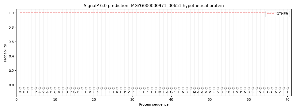
SignalP and Lipop Annotations help
This protein is predicted as OTHER
| Other | SP_Sec_SPI | LIPO_Sec_SPII | TAT_Tat_SPI | TATLIP_Sec_SPII | PILIN_Sec_SPIII |
|---|---|---|---|---|---|
| 1.000046 | 0.000002 | 0.000000 | 0.000000 | 0.000000 | 0.000000 |