You are browsing environment: HUMAN GUT
CAZyme Information: MGYG000000998_00527
You are here: Home > Sequence: MGYG000000998_00527
Basic Information |
Genomic context |
Full Sequence |
Enzyme annotations |
CAZy signature domains |
CDD domains |
CAZyme hits |
PDB hits |
Swiss-Prot hits |
SignalP and Lipop annotations |
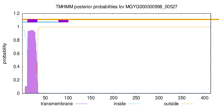
TMHMM annotations
Basic Information help
| Species | TM7x sp900556885 | |||||||||||
|---|---|---|---|---|---|---|---|---|---|---|---|---|
| Lineage | Bacteria; Patescibacteria; Saccharimonadia; Saccharimonadales; Saccharimonadaceae; TM7x; TM7x sp900556885 | |||||||||||
| CAZyme ID | MGYG000000998_00527 | |||||||||||
| CAZy Family | GH16 | |||||||||||
| CAZyme Description | hypothetical protein | |||||||||||
| CAZyme Property |
|
|||||||||||
| Genome Property |
|
|||||||||||
| Gene Location | Start: 11832; End: 13079 Strand: + | |||||||||||
CAZyme Signature Domains help
| Family | Start | End | Evalue | family coverage |
|---|---|---|---|---|
| GH16 | 59 | 310 | 3.5e-67 | 0.9956521739130435 |
CDD Domains download full data without filtering help
| Cdd ID | Domain | E-Value | qStart | qEnd | sStart | sEnd | Domain Description |
|---|---|---|---|---|---|---|---|
| cd08023 | GH16_laminarinase_like | 3.75e-76 | 59 | 310 | 1 | 235 | Laminarinase, member of the glycosyl hydrolase family 16. Laminarinase, also known as glucan endo-1,3-beta-D-glucosidase, is a glycosyl hydrolase family 16 member that hydrolyzes 1,3-beta-D-glucosidic linkages in 1,3-beta-D-glucans such as laminarins, curdlans, paramylons, and pachymans, with very limited action on mixed-link (1,3-1,4-)-beta-D-glucans. |
| cd00413 | Glyco_hydrolase_16 | 1.25e-43 | 61 | 310 | 1 | 210 | glycosyl hydrolase family 16. The O-Glycosyl hydrolases are a widespread group of enzymes that hydrolyse the glycosidic bond between two or more carbohydrates, or between a carbohydrate and a non-carbohydrate moiety. A glycosyl hydrolase classification system based on sequence similarity has led to the definition of more than 95 different families inlcuding glycosyl hydrolase family 16. Family 16 includes lichenase, xyloglucan endotransglycosylase (XET), beta-agarase, kappa-carrageenase, endo-beta-1,3-glucanase, endo-beta-1,3-1,4-glucanase, and endo-beta-galactosidase, all of which have a conserved jelly roll fold with a deep active site channel harboring the catalytic residues. |
| cd08024 | GH16_CCF | 8.16e-43 | 58 | 310 | 2 | 329 | Coelomic cytolytic factor, member of glycosyl hydrolase family 16. Subgroup of glucanases of unknown function that are related to beta-GRP (beta-1,3-glucan recognition protein), but contain active site residues. Beta-GRPs are one group of pattern recognition receptors (PRRs), also referred to as biosensor proteins, that complexes with pathogen-associated beta-1,3-glucans and then transduces signals necessary for activation of an appropriate innate immune response. Beta-GRPs are present in insects and lack all catalytic residues. This subgroup contains related proteins that still contain the active site and are widely distributed in eukaryotes. Their structures adopt a jelly roll fold with a deep active site channel harboring the catalytic residues, like those of other glycosyl hydrolase family 16 members. |
| cd02182 | GH16_Strep_laminarinase_like | 3.34e-40 | 53 | 311 | 2 | 259 | Streptomyces laminarinase-like, member of glycosyl hydrolase family 16. Proteins similar to Streptomyces sioyaensis beta-1,3-glucanase (laminarinase) present in Actinomycetales as well as Peziomycotina. Laminarinases belong to glycosyl hydrolase family 16 and hydrolyze the glycosidic bond of the 1,3-beta-linked glucan, a major component of fungal and plant cell walls and the structural and storage polysaccharides (laminarin) of marine macro-algae. Members of the GH16 family have a conserved jelly roll fold with an active site channel. |
| cd02178 | GH16_beta_agarase | 2.19e-25 | 61 | 310 | 27 | 257 | Beta-agarase, member of glycosyl hydrolase family 16. Beta-agarase is a glycosyl hydrolase family 16 (GH16) member that hydrolyzes the internal beta-1,4-linkage of agarose, a hydrophilic polysaccharide found in the cell wall of Rhodophyceaea, marine red algae. Agarose is a linear chain of galactose units linked by alternating L-alpha-1,3- and D-beta-1,4-linkages that are additionally modified by a 3,6-anhydro-bridge. Agarose forms thermo-reversible gels that are widely used in the food industry or as a laboratory medium. While beta-agarases are also found in two other families derived from the sequence-based classification of glycosyl hydrolases (GH50, and GH86) the GH16 members are most abundant. This domain adopts a curved beta-sandwich conformation, with a tunnel-shaped active site cavity, referred to as a jellyroll fold. |
CAZyme Hits help
| Hit ID | E-Value | Query Start | Query End | Hit Start | Hit End |
|---|---|---|---|---|---|
| QWQ31402.1 | 1.65e-171 | 1 | 411 | 1 | 400 |
| QCT41825.1 | 1.53e-169 | 1 | 411 | 1 | 400 |
| AJA06652.1 | 1.53e-169 | 1 | 411 | 1 | 400 |
| QWQ32251.1 | 5.26e-82 | 66 | 240 | 14 | 191 |
| VEG78372.1 | 1.45e-80 | 52 | 311 | 1455 | 1719 |
PDB Hits download full data without filtering help
| Hit ID | E-Value | Query Start | Query End | Hit Start | Hit End | Description |
|---|---|---|---|---|---|---|
| 5WUT_A | 1.91e-37 | 58 | 311 | 8 | 235 | Crystalstructure of laminarinase from Flavobacterium sp. UMI-01 [Flavobacterium sp.],5WUT_B Crystal structure of laminarinase from Flavobacterium sp. UMI-01 [Flavobacterium sp.] |
| 3ILN_A | 2.91e-37 | 52 | 310 | 4 | 247 | ChainA, Laminarinase [Rhodothermus marinus],3ILN_B Chain B, Laminarinase [Rhodothermus marinus] |
| 2HYK_A | 1.88e-35 | 58 | 311 | 10 | 243 | Thecrystal structure of an endo-beta-1,3-glucanase from alkaliphilic Nocardiopsis sp.strain F96 [Nocardiopsis sp. F96] |
| 4CRQ_A | 1.93e-35 | 58 | 312 | 6 | 232 | Crystalstructure of the catalytic domain of the modular laminarinase ZgLamC mutant E142S [Zobellia galactanivorans],4CRQ_B Crystal structure of the catalytic domain of the modular laminarinase ZgLamC mutant E142S [Zobellia galactanivorans],4CTE_A Crystal structure of the catalytic domain of the modular laminarinase ZgLamC mutant E142S in complex with a thio-oligosaccharide [Zobellia galactanivorans],4CTE_B Crystal structure of the catalytic domain of the modular laminarinase ZgLamC mutant E142S in complex with a thio-oligosaccharide [Zobellia galactanivorans] |
| 2VY0_A | 4.30e-33 | 49 | 310 | 9 | 259 | TheX-ray structure of endo-beta-1,3-glucanase from Pyrococcus furiosus [Pyrococcus furiosus],2VY0_B The X-ray structure of endo-beta-1,3-glucanase from Pyrococcus furiosus [Pyrococcus furiosus] |
Swiss-Prot Hits download full data without filtering help
| Hit ID | E-Value | Query Start | Query End | Hit Start | Hit End | Description |
|---|---|---|---|---|---|---|
| P23903 | 3.86e-44 | 58 | 313 | 426 | 682 | Glucan endo-1,3-beta-glucosidase A1 OS=Niallia circulans OX=1397 GN=glcA PE=1 SV=1 |
| P45798 | 9.89e-38 | 52 | 310 | 39 | 282 | Beta-glucanase OS=Rhodothermus marinus OX=29549 GN=bglA PE=1 SV=1 |
| C1IE32 | 1.89e-35 | 59 | 280 | 24 | 244 | Glucan endo-1,3-beta-glucosidase OS=Cryptopygus antarcticus OX=187623 PE=1 SV=1 |
| Q8N0N3 | 5.18e-31 | 58 | 317 | 33 | 366 | Beta-1,3-glucan-binding protein OS=Penaeus monodon OX=6687 PE=2 SV=1 |
| Q27082 | 4.14e-25 | 58 | 311 | 28 | 253 | Clotting factor G alpha subunit OS=Tachypleus tridentatus OX=6853 PE=1 SV=1 |
SignalP and Lipop Annotations help
This protein is predicted as SP
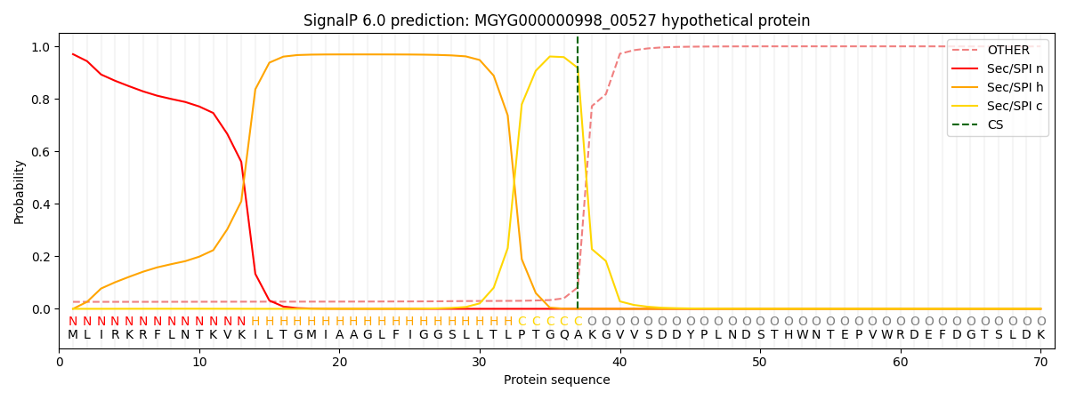
| Other | SP_Sec_SPI | LIPO_Sec_SPII | TAT_Tat_SPI | TATLIP_Sec_SPII | PILIN_Sec_SPIII |
|---|---|---|---|---|---|
| 0.029509 | 0.965572 | 0.003584 | 0.000754 | 0.000281 | 0.000251 |