You are browsing environment: HUMAN GUT
CAZyme Information: MGYG000001087_02672
You are here: Home > Sequence: MGYG000001087_02672
Basic Information |
Genomic context |
Full Sequence |
Enzyme annotations |
CAZy signature domains |
CDD domains |
CAZyme hits |
PDB hits |
Swiss-Prot hits |
SignalP and Lipop annotations |
TMHMM annotations
Basic Information help
| Species | Anaerostipes sp900756035 | |||||||||||
|---|---|---|---|---|---|---|---|---|---|---|---|---|
| Lineage | Bacteria; Firmicutes_A; Clostridia; Lachnospirales; Lachnospiraceae; Anaerostipes; Anaerostipes sp900756035 | |||||||||||
| CAZyme ID | MGYG000001087_02672 | |||||||||||
| CAZy Family | GT4 | |||||||||||
| CAZyme Description | D-inositol-3-phosphate glycosyltransferase | |||||||||||
| CAZyme Property |
|
|||||||||||
| Genome Property |
|
|||||||||||
| Gene Location | Start: 3583; End: 4743 Strand: - | |||||||||||
CDD Domains download full data without filtering help
| Cdd ID | Domain | E-Value | qStart | qEnd | sStart | sEnd | Domain Description |
|---|---|---|---|---|---|---|---|
| COG0438 | RfaB | 1.07e-38 | 1 | 381 | 3 | 380 | Glycosyltransferase involved in cell wall bisynthesis [Cell wall/membrane/envelope biogenesis]. |
| cd03801 | GT4_PimA-like | 1.00e-33 | 27 | 376 | 23 | 366 | phosphatidyl-myo-inositol mannosyltransferase. This family is most closely related to the GT4 family of glycosyltransferases and named after PimA in Propionibacterium freudenreichii, which is involved in the biosynthesis of phosphatidyl-myo-inositol mannosides (PIM) which are early precursors in the biosynthesis of lipomannans (LM) and lipoarabinomannans (LAM), and catalyzes the addition of a mannosyl residue from GDP-D-mannose (GDP-Man) to the position 2 of the carrier lipid phosphatidyl-myo-inositol (PI) to generate a phosphatidyl-myo-inositol bearing an alpha-1,2-linked mannose residue (PIM1). Glycosyltransferases catalyze the transfer of sugar moieties from activated donor molecules to specific acceptor molecules, forming glycosidic bonds. The acceptor molecule can be a lipid, a protein, a heterocyclic compound, or another carbohydrate residue. This group of glycosyltransferases is most closely related to the previously defined glycosyltransferase family 1 (GT1). The members of this family may transfer UDP, ADP, GDP, or CMP linked sugars. The diverse enzymatic activities among members of this family reflect a wide range of biological functions. The protein structure available for this family has the GTB topology, one of the two protein topologies observed for nucleotide-sugar-dependent glycosyltransferases. GTB proteins have distinct N- and C- terminal domains each containing a typical Rossmann fold. The two domains have high structural homology despite minimal sequence homology. The large cleft that separates the two domains includes the catalytic center and permits a high degree of flexibility. The members of this family are found mainly in certain bacteria and archaea. |
| pfam00534 | Glycos_transf_1 | 3.25e-24 | 201 | 353 | 2 | 155 | Glycosyl transferases group 1. Mutations in this domain of PIGA lead to disease (Paroxysmal Nocturnal haemoglobinuria). Members of this family transfer activated sugars to a variety of substrates, including glycogen, Fructose-6-phosphate and lipopolysaccharides. Members of this family transfer UDP, ADP, GDP or CMP linked sugars. The eukaryotic glycogen synthases may be distant members of this family. |
| pfam13692 | Glyco_trans_1_4 | 6.10e-22 | 201 | 344 | 1 | 137 | Glycosyl transferases group 1. |
| cd03811 | GT4_GT28_WabH-like | 8.27e-22 | 27 | 344 | 21 | 327 | family 4 and family 28 glycosyltransferases similar to Klebsiella WabH. This family is most closely related to the GT1 family of glycosyltransferases. WabH in Klebsiella pneumoniae has been shown to transfer a GlcNAc residue from UDP-GlcNAc onto the acceptor GalUA residue in the cellular outer core. |
CAZyme Hits help
| Hit ID | E-Value | Query Start | Query End | Hit Start | Hit End |
|---|---|---|---|---|---|
| ALS18157.1 | 1.20e-132 | 1 | 380 | 1 | 382 |
| CBK98588.1 | 8.08e-123 | 1 | 377 | 1 | 380 |
| AYA78078.1 | 2.54e-74 | 2 | 372 | 4 | 368 |
| AZH31937.1 | 1.15e-71 | 2 | 376 | 4 | 384 |
| AUO05404.1 | 1.62e-71 | 2 | 376 | 4 | 384 |
PDB Hits download full data without filtering help
| Hit ID | E-Value | Query Start | Query End | Hit Start | Hit End | Description |
|---|---|---|---|---|---|---|
| 3C4Q_A | 1.61e-08 | 169 | 383 | 190 | 410 | Structureof the retaining glycosyltransferase MshA : The first step in mycothiol biosynthesis. Organism : Corynebacterium glutamicum- Complex with UDP [Corynebacterium glutamicum],3C4Q_B Structure of the retaining glycosyltransferase MshA : The first step in mycothiol biosynthesis. Organism : Corynebacterium glutamicum- Complex with UDP [Corynebacterium glutamicum],3C4V_A Structure of the retaining glycosyltransferase MshA:The first step in mycothiol biosynthesis. Organism: Corynebacterium glutamicum : Complex with UDP and 1L-INS-1-P. [Corynebacterium glutamicum],3C4V_B Structure of the retaining glycosyltransferase MshA:The first step in mycothiol biosynthesis. Organism: Corynebacterium glutamicum : Complex with UDP and 1L-INS-1-P. [Corynebacterium glutamicum] |
| 3C48_A | 1.65e-08 | 169 | 383 | 210 | 430 | Structureof the retaining glycosyltransferase MshA: The first step in mycothiol biosynthesis. Organism: Corynebacterium glutamicum- APO (OPEN) structure. [Corynebacterium glutamicum],3C48_B Structure of the retaining glycosyltransferase MshA: The first step in mycothiol biosynthesis. Organism: Corynebacterium glutamicum- APO (OPEN) structure. [Corynebacterium glutamicum] |
| 6D9T_A | 3.56e-08 | 199 | 384 | 215 | 396 | BshAfrom Staphylococcus aureus complexed with UDP [Staphylococcus aureus] |
| 6N1X_A | 1.05e-07 | 199 | 380 | 199 | 376 | ChainA, Glycosyltransferase [Staphylococcus aureus subsp. aureus CN1] |
| 5D00_A | 1.06e-07 | 199 | 378 | 200 | 375 | Crystalstructure of BshA from B. subtilis complexed with N-acetylglucosaminyl-malate and UMP [Bacillus subtilis subsp. subtilis str. 168],5D00_B Crystal structure of BshA from B. subtilis complexed with N-acetylglucosaminyl-malate and UMP [Bacillus subtilis subsp. subtilis str. 168],5D01_A Crystal structure of BshA from B. subtilis complexed with N-acetylglucosaminyl-malate [Bacillus subtilis subsp. subtilis str. 168],5D01_B Crystal structure of BshA from B. subtilis complexed with N-acetylglucosaminyl-malate [Bacillus subtilis subsp. subtilis str. 168] |
Swiss-Prot Hits download full data without filtering help
| Hit ID | E-Value | Query Start | Query End | Hit Start | Hit End | Description |
|---|---|---|---|---|---|---|
| A0QQZ8 | 5.01e-14 | 155 | 375 | 187 | 403 | D-inositol 3-phosphate glycosyltransferase OS=Mycolicibacterium smegmatis (strain ATCC 700084 / mc(2)155) OX=246196 GN=mshA PE=1 SV=1 |
| Q59002 | 5.53e-14 | 196 | 376 | 204 | 382 | Uncharacterized glycosyltransferase MJ1607 OS=Methanocaldococcus jannaschii (strain ATCC 43067 / DSM 2661 / JAL-1 / JCM 10045 / NBRC 100440) OX=243232 GN=MJ1607 PE=3 SV=1 |
| Q4JSW2 | 1.14e-13 | 156 | 378 | 177 | 406 | D-inositol 3-phosphate glycosyltransferase OS=Corynebacterium jeikeium (strain K411) OX=306537 GN=mshA PE=3 SV=1 |
| Q48453 | 3.61e-13 | 149 | 376 | 141 | 356 | Uncharacterized 41.2 kDa protein in cps region OS=Klebsiella pneumoniae OX=573 PE=4 SV=1 |
| D6Z995 | 3.06e-12 | 204 | 376 | 258 | 433 | D-inositol 3-phosphate glycosyltransferase OS=Segniliparus rotundus (strain ATCC BAA-972 / CDC 1076 / CIP 108378 / DSM 44985 / JCM 13578) OX=640132 GN=mshA PE=3 SV=1 |
SignalP and Lipop Annotations help
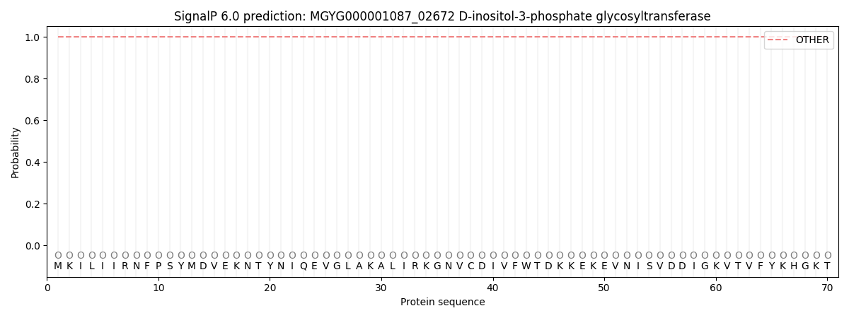
This protein is predicted as OTHER
| Other | SP_Sec_SPI | LIPO_Sec_SPII | TAT_Tat_SPI | TATLIP_Sec_SPII | PILIN_Sec_SPIII |
|---|---|---|---|---|---|
| 1.000031 | 0.000000 | 0.000000 | 0.000000 | 0.000000 | 0.000000 |