You are browsing environment: HUMAN GUT
CAZyme Information: MGYG000001149_01069
You are here: Home > Sequence: MGYG000001149_01069
Basic Information |
Genomic context |
Full Sequence |
Enzyme annotations |
CAZy signature domains |
CDD domains |
CAZyme hits |
PDB hits |
Swiss-Prot hits |
SignalP and Lipop annotations |
TMHMM annotations
Basic Information help
| Species | Fusobacterium_B sp900554355 | |||||||||||
|---|---|---|---|---|---|---|---|---|---|---|---|---|
| Lineage | Bacteria; Fusobacteriota; Fusobacteriia; Fusobacteriales; Fusobacteriaceae; Fusobacterium_B; Fusobacterium_B sp900554355 | |||||||||||
| CAZyme ID | MGYG000001149_01069 | |||||||||||
| CAZy Family | CE4 | |||||||||||
| CAZyme Description | hypothetical protein | |||||||||||
| CAZyme Property |
|
|||||||||||
| Genome Property |
|
|||||||||||
| Gene Location | Start: 53574; End: 55379 Strand: - | |||||||||||
CAZyme Signature Domains help
| Family | Start | End | Evalue | family coverage |
|---|---|---|---|---|
| CE4 | 409 | 550 | 1.3e-31 | 0.9461538461538461 |
CDD Domains download full data without filtering help
| Cdd ID | Domain | E-Value | qStart | qEnd | sStart | sEnd | Domain Description |
|---|---|---|---|---|---|---|---|
| cd03819 | GT4_WavL-like | 3.96e-62 | 3 | 327 | 1 | 343 | Vibrio cholerae WavL and similar sequences. This family is most closely related to the GT4 family of glycosyltransferases. WavL in Vibrio cholerae has been shown to be involved in the biosynthesis of the lipopolysaccharide core. |
| cd10918 | CE4_NodB_like_5s_6s | 3.91e-55 | 415 | 572 | 2 | 157 | Putative catalytic NodB homology domain of PgaB, IcaB, and similar proteins which consist of a deformed (beta/alpha)8 barrel fold with 5- or 6-strands. This family belongs to the large and functionally diverse carbohydrate esterase 4 (CE4) superfamily, whose members show strong sequence similarity with some variability due to their distinct carbohydrate substrates. It includes bacterial poly-beta-1,6-N-acetyl-D-glucosamine N-deacetylase PgaB, hemin storage system HmsF protein in gram-negative species, intercellular adhesion proteins IcaB, and many uncharacterized prokaryotic polysaccharide deacetylases. It also includes a putative polysaccharide deacetylase YxkH encoded by the Bacillus subtilis yxkH gene, which is one of six polysaccharide deacetylase gene homologs present in the Bacillus subtilis genome. Sequence comparison shows all family members contain a conserved domain similar to the catalytic NodB homology domain of rhizobial NodB-like proteins, which consists of a deformed (beta/alpha)8 barrel fold with 6 or 7 strands. However, in this family, most proteins have 5 strands and some have 6 strands. Moreover, long insertions are found in many family members, whose function remains unknown. |
| cd10969 | CE4_Ecf1_like_5s | 5.99e-45 | 377 | 581 | 2 | 213 | Putative catalytic NodB homology domain of a hypothetical protein Ecf1 from Escherichia coli and similar proteins. This family contains a hypothetical protein Ecf1 from Escherichia coli and its prokaryotic homologs. Although their biochemical properties remain to be determined, members in this family contain a conserved domain with a 5-stranded beta/alpha barrel, which is similar to the catalytic NodB homology domain of rhizobial NodB-like proteins, belonging to the larger carbohydrate esterase 4 (CE4) superfamily. |
| cd10973 | CE4_DAC_u4_5s | 8.74e-37 | 413 | 575 | 1 | 156 | Putative catalytic NodB homology domain of uncharacterized bacterial polysaccharide deacetylases which consist of a 5-stranded beta/alpha barrel. This family contains many uncharacterized bacterial polysaccharide deacetylases. Although their biological functions remain unknown, all members of the family are predicted to contain a conserved domain with a 5-stranded beta/alpha barrel, which is similar to the catalytic NodB homology domain of rhizobial NodB-like proteins, belonging to the larger carbohydrate esterase 4 (CE4) superfamily. |
| cd03801 | GT4_PimA-like | 1.65e-31 | 2 | 339 | 1 | 365 | phosphatidyl-myo-inositol mannosyltransferase. This family is most closely related to the GT4 family of glycosyltransferases and named after PimA in Propionibacterium freudenreichii, which is involved in the biosynthesis of phosphatidyl-myo-inositol mannosides (PIM) which are early precursors in the biosynthesis of lipomannans (LM) and lipoarabinomannans (LAM), and catalyzes the addition of a mannosyl residue from GDP-D-mannose (GDP-Man) to the position 2 of the carrier lipid phosphatidyl-myo-inositol (PI) to generate a phosphatidyl-myo-inositol bearing an alpha-1,2-linked mannose residue (PIM1). Glycosyltransferases catalyze the transfer of sugar moieties from activated donor molecules to specific acceptor molecules, forming glycosidic bonds. The acceptor molecule can be a lipid, a protein, a heterocyclic compound, or another carbohydrate residue. This group of glycosyltransferases is most closely related to the previously defined glycosyltransferase family 1 (GT1). The members of this family may transfer UDP, ADP, GDP, or CMP linked sugars. The diverse enzymatic activities among members of this family reflect a wide range of biological functions. The protein structure available for this family has the GTB topology, one of the two protein topologies observed for nucleotide-sugar-dependent glycosyltransferases. GTB proteins have distinct N- and C- terminal domains each containing a typical Rossmann fold. The two domains have high structural homology despite minimal sequence homology. The large cleft that separates the two domains includes the catalytic center and permits a high degree of flexibility. The members of this family are found mainly in certain bacteria and archaea. |
CAZyme Hits help
| Hit ID | E-Value | Query Start | Query End | Hit Start | Hit End |
|---|---|---|---|---|---|
| AVQ29065.1 | 3.82e-265 | 1 | 598 | 1 | 598 |
| SQJ02331.1 | 3.82e-265 | 1 | 598 | 1 | 598 |
| VEH39037.1 | 1.95e-260 | 1 | 598 | 1 | 598 |
| AVQ32077.1 | 1.95e-260 | 1 | 598 | 1 | 598 |
| BBA51388.1 | 2.76e-260 | 1 | 598 | 1 | 598 |
PDB Hits download full data without filtering help
| Hit ID | E-Value | Query Start | Query End | Hit Start | Hit End | Description |
|---|---|---|---|---|---|---|
| 6DQ3_A | 9.87e-20 | 350 | 571 | 8 | 217 | ChainA, Polysaccharide deacetylase [Streptococcus pyogenes],6DQ3_B Chain B, Polysaccharide deacetylase [Streptococcus pyogenes] |
| 4V33_A | 5.84e-19 | 350 | 578 | 143 | 355 | Crystalstructure of the putative polysaccharide deacetylase BA0330 from bacillus anthracis [Bacillus anthracis],4V33_B Crystal structure of the putative polysaccharide deacetylase BA0330 from bacillus anthracis [Bacillus anthracis] |
| 4HD5_A | 1.41e-18 | 350 | 578 | 143 | 355 | CrystalStructure of BC0361, a polysaccharide deacetylase from Bacillus cereus [Bacillus cereus ATCC 14579] |
| 4U10_A | 1.27e-09 | 353 | 579 | 6 | 260 | Probingthe structure and mechanism of de-N-acetylase from aggregatibacter actinomycetemcomitans [Aggregatibacter actinomycetemcomitans],4U10_B Probing the structure and mechanism of de-N-acetylase from aggregatibacter actinomycetemcomitans [Aggregatibacter actinomycetemcomitans] |
| 4WCJ_A | 4.92e-08 | 353 | 544 | 39 | 224 | Structureof IcaB from Ammonifex degensii [Ammonifex degensii KC4] |
Swiss-Prot Hits download full data without filtering help
| Hit ID | E-Value | Query Start | Query End | Hit Start | Hit End | Description |
|---|---|---|---|---|---|---|
| P94361 | 8.33e-27 | 346 | 571 | 60 | 264 | Putative polysaccharide deacetylase YxkH OS=Bacillus subtilis (strain 168) OX=224308 GN=yxkH PE=3 SV=1 |
| P31666 | 2.28e-14 | 351 | 583 | 170 | 404 | Uncharacterized protein YadE OS=Escherichia coli (strain K12) OX=83333 GN=yadE PE=3 SV=2 |
| P75906 | 1.86e-06 | 354 | 578 | 52 | 301 | Poly-beta-1,6-N-acetyl-D-glucosamine N-deacetylase OS=Escherichia coli (strain K12) OX=83333 GN=pgaB PE=1 SV=1 |
| Q8XAR3 | 2.45e-06 | 354 | 578 | 52 | 301 | Poly-beta-1,6-N-acetyl-D-glucosamine N-deacetylase OS=Escherichia coli O157:H7 OX=83334 GN=pgaB PE=3 SV=1 |
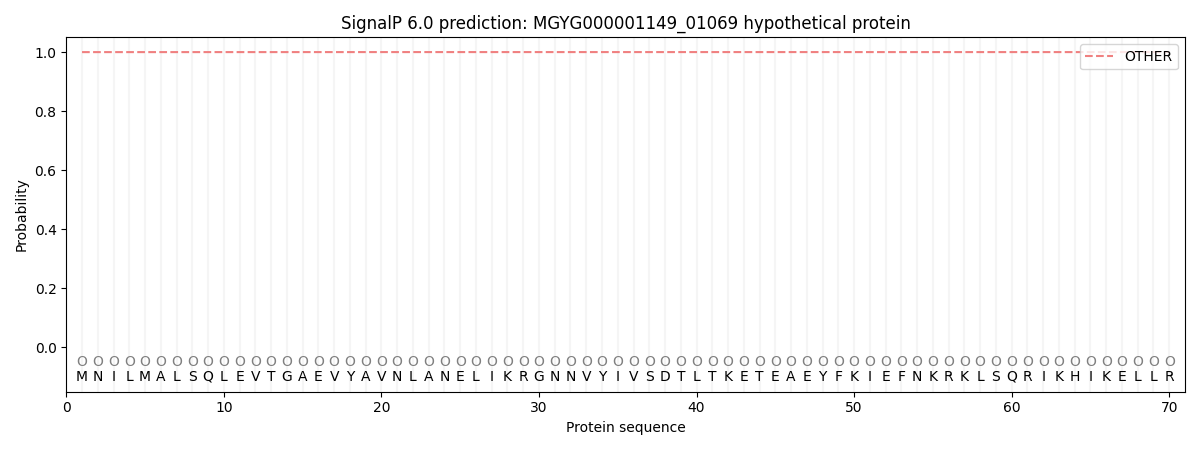
SignalP and Lipop Annotations help
This protein is predicted as OTHER
| Other | SP_Sec_SPI | LIPO_Sec_SPII | TAT_Tat_SPI | TATLIP_Sec_SPII | PILIN_Sec_SPIII |
|---|---|---|---|---|---|
| 1.000030 | 0.000004 | 0.000000 | 0.000000 | 0.000000 | 0.000000 |