You are browsing environment: HUMAN GUT
CAZyme Information: MGYG000001195_00697
You are here: Home > Sequence: MGYG000001195_00697
Basic Information |
Genomic context |
Full Sequence |
Enzyme annotations |
CAZy signature domains |
CDD domains |
CAZyme hits |
PDB hits |
Swiss-Prot hits |
SignalP and Lipop annotations |
TMHMM annotations
Basic Information help
| Species | ||||||||||||
|---|---|---|---|---|---|---|---|---|---|---|---|---|
| Lineage | Bacteria; Firmicutes_A; Clostridia; TANB77; CAG-508; UMGS1994; | |||||||||||
| CAZyme ID | MGYG000001195_00697 | |||||||||||
| CAZy Family | GT81 | |||||||||||
| CAZyme Description | Glucosyl-3-phosphoglycerate synthase | |||||||||||
| CAZyme Property |
|
|||||||||||
| Genome Property |
|
|||||||||||
| Gene Location | Start: 131244; End: 131894 Strand: + | |||||||||||
CAZyme Signature Domains help
| Family | Start | End | Evalue | family coverage |
|---|---|---|---|---|
| GT81 | 3 | 213 | 3e-52 | 0.7986348122866894 |
CDD Domains download full data without filtering help
| Cdd ID | Domain | E-Value | qStart | qEnd | sStart | sEnd | Domain Description |
|---|---|---|---|---|---|---|---|
| PRK13915 | PRK13915 | 3.61e-53 | 4 | 213 | 33 | 267 | putative glucosyl-3-phosphoglycerate synthase; Provisional |
| cd04179 | DPM_DPG-synthase_like | 1.07e-27 | 6 | 122 | 1 | 125 | DPM_DPG-synthase_like is a member of the Glycosyltransferase 2 superfamily. DPM1 is the catalytic subunit of eukaryotic dolichol-phosphate mannose (DPM) synthase. DPM synthase is required for synthesis of the glycosylphosphatidylinositol (GPI) anchor, N-glycan precursor, protein O-mannose, and C-mannose. In higher eukaryotes,the enzyme has three subunits, DPM1, DPM2 and DPM3. DPM is synthesized from dolichol phosphate and GDP-Man on the cytosolic surface of the ER membrane by DPM synthase and then is flipped onto the luminal side and used as a donor substrate. In lower eukaryotes, such as Saccharomyces cerevisiae and Trypanosoma brucei, DPM synthase consists of a single component (Dpm1p and TbDpm1, respectively) that possesses one predicted transmembrane region near the C terminus for anchoring to the ER membrane. In contrast, the Dpm1 homologues of higher eukaryotes, namely fission yeast, fungi, and animals, have no transmembrane region, suggesting the existence of adapter molecules for membrane anchoring. This family also includes bacteria and archaea DPM1_like enzymes. However, the enzyme structure and mechanism of function are not well understood. The UDP-glucose:dolichyl-phosphate glucosyltransferase (DPG_synthase) is a transmembrane-bound enzyme of the endoplasmic reticulum involved in protein N-linked glycosylation. This enzyme catalyzes the transfer of glucose from UDP-glucose to dolichyl phosphate. This protein family belongs to Glycosyltransferase 2 superfamily. |
| pfam00535 | Glycos_transf_2 | 4.02e-24 | 5 | 93 | 1 | 96 | Glycosyl transferase family 2. Diverse family, transferring sugar from UDP-glucose, UDP-N-acetyl- galactosamine, GDP-mannose or CDP-abequose, to a range of substrates including cellulose, dolichol phosphate and teichoic acids. |
| COG0463 | WcaA | 1.70e-23 | 1 | 94 | 2 | 102 | Glycosyltransferase involved in cell wall bisynthesis [Cell wall/membrane/envelope biogenesis]. |
| cd00761 | Glyco_tranf_GTA_type | 5.74e-23 | 6 | 140 | 1 | 151 | Glycosyltransferase family A (GT-A) includes diverse families of glycosyl transferases with a common GT-A type structural fold. Glycosyltransferases (GTs) are enzymes that synthesize oligosaccharides, polysaccharides, and glycoconjugates by transferring the sugar moiety from an activated nucleotide-sugar donor to an acceptor molecule, which may be a growing oligosaccharide, a lipid, or a protein. Based on the stereochemistry of the donor and acceptor molecules, GTs are classified as either retaining or inverting enzymes. To date, all GT structures adopt one of two possible folds, termed GT-A fold and GT-B fold. This hierarchy includes diverse families of glycosyl transferases with a common GT-A type structural fold, which has two tightly associated beta/alpha/beta domains that tend to form a continuous central sheet of at least eight beta-strands. The majority of the proteins in this superfamily are Glycosyltransferase family 2 (GT-2) proteins. But it also includes families GT-43, GT-6, GT-8, GT13 and GT-7; which are evolutionarily related to GT-2 and share structure similarities. |
CAZyme Hits help
| Hit ID | E-Value | Query Start | Query End | Hit Start | Hit End |
|---|---|---|---|---|---|
| ASZ10007.1 | 8.48e-66 | 4 | 213 | 7 | 216 |
| QEH40387.1 | 5.88e-64 | 4 | 213 | 2 | 211 |
| QJB41308.1 | 1.47e-63 | 4 | 213 | 2 | 211 |
| QJB34794.1 | 1.47e-63 | 4 | 213 | 2 | 211 |
| AWO01786.1 | 1.51e-63 | 4 | 213 | 25 | 234 |
PDB Hits download full data without filtering help
| Hit ID | E-Value | Query Start | Query End | Hit Start | Hit End | Description |
|---|---|---|---|---|---|---|
| 3CKJ_A | 1.40e-29 | 2 | 212 | 48 | 285 | CrystalStructure of a Mycobacterial Protein [Mycobacterium avium subsp. paratuberculosis],3CKN_A Crystal Structure of a Mycobacterial Protein [Mycobacterium avium subsp. paratuberculosis],3CKO_A Crystal Structure of a Mycobacterial Protein [Mycobacterium avium subsp. paratuberculosis],3CKQ_A Crystal Structure of a Mycobacterial Protein [Mycobacterium avium subsp. paratuberculosis],3CKV_A Crystal Structure of a Mycobacterial Protein [Mycobacterium avium subsp. paratuberculosis] |
| 4Y6N_A | 7.32e-29 | 2 | 212 | 47 | 284 | Crystalstructure of glucosyl-3-phosphoglycerate synthase from Mycobacterium tuberculosis in complex with Mn2+, uridine-diphosphate-glucose (UDP-Glc) and phosphoglyceric acid (PGA) - GpgS Mn2+ UDP-Glc PGA-1 [Mycobacterium tuberculosis H37Rv],4Y6U_A Mycobacterial protein [Mycobacterium tuberculosis H37Rv],4Y7F_A Crystal structure of glucosyl-3-phosphoglycerate synthase from Mycobacterium tuberculosis in complex with Mn2+, uridine-diphosphate-glucose (UDP-Glc) and 3-(phosphonooxy)propanoic acid (PPA) - GpgS Mn2+ UDP-Glc PPA [Mycobacterium tuberculosis H37Rv],4Y7G_A Crystal structure of glucosyl-3-phosphoglycerate synthase from Mycobacterium tuberculosis in complex with Mn2+, uridine-diphosphate-glucose (UDP-Glc) and glycerol 3-phosphate (G3P) - GpgS Mn2+ UDP-Glc G3P [Mycobacterium tuberculosis H37Rv],4Y9X_A Crystal structure of glucosyl-3-phosphoglycerate synthase from Mycobacterium tuberculosis in complex with Mn2+, uridine-diphosphate-glucose (UDP-Glc) and phosphoglyceric acid (PGA) - GpgS Mn2+ UDP-Glc PGA-3 [Mycobacterium tuberculosis H37Rv],5JQX_A Crystal structure of glucosyl-3-phosphoglycerate synthase from Mycobacterium tuberculosis in complex with phosphoglyceric acid (PGA) - GpgS*PGA [Mycobacterium tuberculosis H37Ra],5JQX_B Crystal structure of glucosyl-3-phosphoglycerate synthase from Mycobacterium tuberculosis in complex with phosphoglyceric acid (PGA) - GpgS*PGA [Mycobacterium tuberculosis H37Ra],5JQX_C Crystal structure of glucosyl-3-phosphoglycerate synthase from Mycobacterium tuberculosis in complex with phosphoglyceric acid (PGA) - GpgS*PGA [Mycobacterium tuberculosis H37Ra],5JQX_D Crystal structure of glucosyl-3-phosphoglycerate synthase from Mycobacterium tuberculosis in complex with phosphoglyceric acid (PGA) - GpgS*PGA [Mycobacterium tuberculosis H37Ra],5JSX_A Crystal structure of glucosyl-3-phosphoglycerate synthase from Mycobacterium tuberculosis in complex with Mn2+ and uridine-diphosphate-glucose (UDP-Glc) [Mycobacterium tuberculosis H37Ra],5JT0_A Crystal structure of glucosyl-3-phosphoglycerate synthase from Mycobacterium tuberculosis in complex with Mn2+, uridine-diphosphate (UDP) and glucosyl-3-phosphoglycerate (GPG) - GpgS*GPG*UDP*Mn2+ [Mycobacterium tuberculosis H37Rv],5JUC_A Crystal structure of glucosyl-3-phosphoglycerate synthase from Mycobacterium tuberculosis in complex with Mn2+, uridine-diphosphate (UDP) and glucosyl-3-phosphoglycerate (GPG) - GpgS*GPG*UDP*Mn2+_2 [Mycobacterium tuberculosis H37Rv],5JUD_A Crystal structure of glucosyl-3-phosphoglycerate synthase from Mycobacterium tuberculosis in complex with uridine-diphosphate (UDP) - GpgS*UDP [Mycobacterium tuberculosis variant bovis AF2122/97] |
| 3E25_A | 8.51e-29 | 2 | 212 | 43 | 280 | ChainA, Crystal structure of M. tuberculosis glucosyl-3-phosphoglycerate synthase [Mycobacterium tuberculosis],3E26_A Chain A, Crystal structure of M. tuberculosis glucosyl-3-phosphoglycerate synthase [Mycobacterium tuberculosis] |
| 4DDZ_A | 9.53e-29 | 2 | 212 | 63 | 300 | Crystalstructure of glucosyl-3-phosphoglycerate synthase from Mycobacterium tuberculosis [Mycobacterium tuberculosis H37Rv],4DE7_A Crystal structure of glucosyl-3-phosphoglycerate synthase from Mycobacterium tuberculosis in complex with Mg2+ and uridine-diphosphate (UDP) [Mycobacterium tuberculosis H37Rv],4DEC_A Crystal structure of glucosyl-3-phosphoglycerate synthase from Mycobacterium tuberculosis in complex with Mn2+, uridine-diphosphate (UDP) and phosphoglyceric acid (PGA) [Mycobacterium tuberculosis H37Rv],5JQQ_A Crystal structure of glucosyl-3-phosphoglycerate synthase from Mycobacterium tuberculosis - apo form [Mycobacterium tuberculosis H37Ra] |
| 3F1Y_A | 2.46e-27 | 4 | 214 | 96 | 332 | Mannosyl-3-phosphoglyceratesynthase from Rubrobacter xylanophilus [synthetic construct],3F1Y_C Mannosyl-3-phosphoglycerate synthase from Rubrobacter xylanophilus [synthetic construct],3KIA_A Crystal structure of mannosyl-3-phosphoglycerate synthase from Rubrobacter xylanophilus [synthetic construct],3KIA_C Crystal structure of mannosyl-3-phosphoglycerate synthase from Rubrobacter xylanophilus [synthetic construct],3O3P_A Crystal structure of R. xylanophilus MpgS in complex with GDP-Mannose [Rubrobacter xylanophilus],3O3P_B Crystal structure of R. xylanophilus MpgS in complex with GDP-Mannose [Rubrobacter xylanophilus] |
Swiss-Prot Hits download full data without filtering help
| Hit ID | E-Value | Query Start | Query End | Hit Start | Hit End | Description |
|---|---|---|---|---|---|---|
| Q73WU1 | 7.67e-29 | 2 | 212 | 48 | 285 | Glucosyl-3-phosphoglycerate synthase OS=Mycolicibacterium paratuberculosis (strain ATCC BAA-968 / K-10) OX=262316 GN=MAP_2569c PE=1 SV=1 |
| P9WMW9 | 3.74e-28 | 2 | 212 | 43 | 280 | Glucosyl-3-phosphoglycerate synthase OS=Mycobacterium tuberculosis (strain ATCC 25618 / H37Rv) OX=83332 GN=gpgS PE=1 SV=1 |
| Q7U0E1 | 3.74e-28 | 2 | 212 | 43 | 280 | Glucosyl-3-phosphoglycerate synthase OS=Mycobacterium bovis (strain ATCC BAA-935 / AF2122/97) OX=233413 GN=gpgS PE=1 SV=1 |
| P9WMW8 | 3.74e-28 | 2 | 212 | 43 | 280 | Glucosyl-3-phosphoglycerate synthase OS=Mycobacterium tuberculosis (strain CDC 1551 / Oshkosh) OX=83331 GN=gpgS PE=3 SV=1 |
| A0R2E6 | 3.75e-27 | 2 | 212 | 28 | 262 | Glucosyl-3-phosphoglycerate synthase OS=Mycolicibacterium smegmatis (strain ATCC 700084 / mc(2)155) OX=246196 GN=gpgS PE=1 SV=1 |
SignalP and Lipop Annotations help
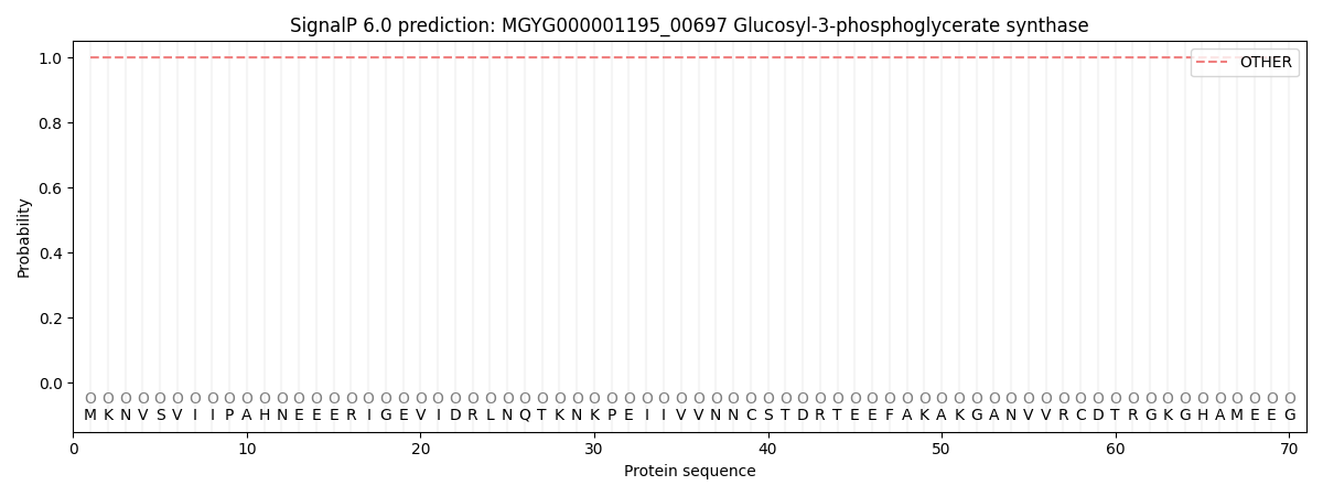
This protein is predicted as OTHER
| Other | SP_Sec_SPI | LIPO_Sec_SPII | TAT_Tat_SPI | TATLIP_Sec_SPII | PILIN_Sec_SPIII |
|---|---|---|---|---|---|
| 1.000064 | 0.000000 | 0.000000 | 0.000000 | 0.000000 | 0.000000 |