You are browsing environment: HUMAN GUT
CAZyme Information: MGYG000001217_01051
You are here: Home > Sequence: MGYG000001217_01051
Basic Information |
Genomic context |
Full Sequence |
Enzyme annotations |
CAZy signature domains |
CDD domains |
CAZyme hits |
PDB hits |
Swiss-Prot hits |
SignalP and Lipop annotations |
TMHMM annotations
Basic Information help
| Species | Firm-10 sp004552515 | |||||||||||
|---|---|---|---|---|---|---|---|---|---|---|---|---|
| Lineage | Bacteria; Firmicutes_A; Clostridia_A; Christensenellales; CAG-74; Firm-10; Firm-10 sp004552515 | |||||||||||
| CAZyme ID | MGYG000001217_01051 | |||||||||||
| CAZy Family | GH4 | |||||||||||
| CAZyme Description | Alpha-galactosidase | |||||||||||
| CAZyme Property |
|
|||||||||||
| Genome Property |
|
|||||||||||
| Gene Location | Start: 373; End: 1695 Strand: - | |||||||||||
CAZyme Signature Domains help
| Family | Start | End | Evalue | family coverage |
|---|---|---|---|---|
| GH4 | 4 | 183 | 1.1e-48 | 0.9888268156424581 |
CDD Domains download full data without filtering help
| Cdd ID | Domain | E-Value | qStart | qEnd | sStart | sEnd | Domain Description |
|---|---|---|---|---|---|---|---|
| PRK15076 | PRK15076 | 1.51e-169 | 4 | 439 | 3 | 431 | alpha-galactosidase; Provisional |
| cd05297 | GH4_alpha_glucosidase_galactosidase | 1.46e-159 | 4 | 428 | 2 | 423 | Glycoside Hydrolases Family 4; Alpha-glucosidases and alpha-galactosidases. linked to 3D####ucture |
| COG1486 | CelF | 1.47e-113 | 1 | 439 | 2 | 440 | Alpha-galactosidase/6-phospho-beta-glucosidase, family 4 of glycosyl hydrolase [Carbohydrate transport and metabolism]. |
| cd05296 | GH4_P_beta_glucosidase | 7.19e-48 | 3 | 435 | 1 | 419 | Glycoside Hydrolases Family 4; Phospho-beta-glucosidase. Some bacteria simultaneously translocate and phosphorylate disaccharides via the phosphoenolpyruvate-dependent phosphotransferase system (PEP-PTS). After translocation, these phospho-disaccharides may be hydrolyzed by the GH4 glycoside hydrolases such as the phospho-beta-glucosidases. Other organisms (such as archaea and Thermotoga maritima ) lack the PEP-PTS system, but have several enzymes normally associated with the PEP-PTS operon. The 6-phospho-beta-glucosidase from Thermotoga maritima hydrolylzes cellobiose 6-phosphate (6P) into glucose-6P and glucose, in an NAD+ and Mn2+ dependent fashion. The Escherichia coli 6-phospho-beta-glucosidase (also called celF) hydrolyzes a variety of phospho-beta-glucosides including cellobiose-6P, salicin-6P, arbutin-6P, and gentobiose-6P. Phospho-beta-glucosidases are part of the NAD(P)-binding Rossmann fold superfamily, which includes a wide variety of protein families including the NAD(P)-binding domains of alcohol dehydrogenases, tyrosine-dependent oxidoreductases, glyceraldehyde-3-phosphate dehydrogenases, formate/glycerate dehydrogenases, siroheme synthases, 6-phosphogluconate dehydrogenases, aminoacid dehydrogenases, repressor rex, and NAD-binding potassium channel domains, among others. |
| cd05197 | GH4_glycoside_hydrolases | 5.77e-47 | 4 | 429 | 2 | 424 | Glycoside Hydrases Family 4. Glycoside hydrolases cleave glycosidic bonds to release smaller sugars from oligo- or polysaccharides. Some bacteria simultaneously translocate and phosphorylate disaccharides via the phosphoenolpyruvate-dependent phosphotransferase system (PEP-PTS). After translocation, these phospho-disaccharides may be hydrolyzed by GH4 glycoside hydrolases. Other organisms (such as archaea and Thermotoga maritima) lack the PEP-PTS system, but have several enzymes normally associated with the PEP-PTS operon. GH4 family members include 6-phospho-beta-glucosidases, 6-phospho-alpha-glucosidases, alpha-glucosidases/alpha-glucuronidases (only from Thermotoga), and alpha-galactosidases. They require two cofactors, NAD+ and a divalent metal (Mn2+, Ni2+, Mg2+), for activity. Some also require reducing conditions. GH4 glycoside hydrolases are part of the NAD(P)-binding Rossmann fold superfamily, which includes a wide variety of protein families including the NAD(P)-binding domains of alcohol dehydrogenases, tyrosine-dependent oxidoreductases, glyceraldehyde-3-phosphate dehydrogenases, formate/glycerate dehydrogenases, siroheme synthases, 6-phosphogluconate dehydrogenases, aminoacid dehydrogenases, repressor rex, and NAD-binding potassium channel domains, among others. |
CAZyme Hits help
| Hit ID | E-Value | Query Start | Query End | Hit Start | Hit End |
|---|---|---|---|---|---|
| ANE45532.1 | 1.14e-177 | 4 | 438 | 3 | 436 |
| AUS96821.1 | 2.37e-177 | 3 | 440 | 2 | 438 |
| QHT59435.1 | 3.16e-175 | 1 | 439 | 1 | 439 |
| QHT62813.1 | 4.65e-175 | 3 | 439 | 2 | 436 |
| QHW31407.1 | 1.19e-174 | 1 | 439 | 1 | 439 |
PDB Hits download full data without filtering help
| Hit ID | E-Value | Query Start | Query End | Hit Start | Hit End | Description |
|---|---|---|---|---|---|---|
| 5C3M_A | 4.23e-39 | 4 | 438 | 6 | 437 | Crystalstructure of Gan4C, a GH4 6-phospho-glucosidase from Geobacillus stearothermophilus [Geobacillus stearothermophilus],5C3M_B Crystal structure of Gan4C, a GH4 6-phospho-glucosidase from Geobacillus stearothermophilus [Geobacillus stearothermophilus],5C3M_C Crystal structure of Gan4C, a GH4 6-phospho-glucosidase from Geobacillus stearothermophilus [Geobacillus stearothermophilus],5C3M_D Crystal structure of Gan4C, a GH4 6-phospho-glucosidase from Geobacillus stearothermophilus [Geobacillus stearothermophilus] |
| 1S6Y_A | 3.89e-36 | 4 | 438 | 9 | 440 | 2.3Acrystal structure of phospho-beta-glucosidase [Geobacillus stearothermophilus] |
| 3FEF_A | 7.39e-36 | 4 | 410 | 7 | 420 | Crystalstructure of putative glucosidase lplD from bacillus subtilis [Bacillus subtilis],3FEF_B Crystal structure of putative glucosidase lplD from bacillus subtilis [Bacillus subtilis],3FEF_C Crystal structure of putative glucosidase lplD from bacillus subtilis [Bacillus subtilis],3FEF_D Crystal structure of putative glucosidase lplD from bacillus subtilis [Bacillus subtilis] |
| 1U8X_X | 2.52e-19 | 6 | 437 | 31 | 463 | CrystalStructure Of Glva From Bacillus Subtilis, A Metal-requiring, Nad-dependent 6-phospho-alpha-glucosidase [Bacillus subtilis] |
| 6VC6_A | 2.87e-19 | 8 | 439 | 9 | 441 | 2.1Angstrom Resolution Crystal Structure of 6-phospho-alpha-glucosidase from Gut Microorganisms in Complex with NAD and Mn2+ [Merdibacter massiliensis],6VC6_B 2.1 Angstrom Resolution Crystal Structure of 6-phospho-alpha-glucosidase from Gut Microorganisms in Complex with NAD and Mn2+ [Merdibacter massiliensis],6VC6_C 2.1 Angstrom Resolution Crystal Structure of 6-phospho-alpha-glucosidase from Gut Microorganisms in Complex with NAD and Mn2+ [Merdibacter massiliensis],6VC6_D 2.1 Angstrom Resolution Crystal Structure of 6-phospho-alpha-glucosidase from Gut Microorganisms in Complex with NAD and Mn2+ [Merdibacter massiliensis] |
Swiss-Prot Hits download full data without filtering help
| Hit ID | E-Value | Query Start | Query End | Hit Start | Hit End | Description |
|---|---|---|---|---|---|---|
| P06720 | 1.07e-95 | 4 | 436 | 6 | 446 | Alpha-galactosidase OS=Escherichia coli (strain K12) OX=83333 GN=melA PE=1 SV=1 |
| P30877 | 4.24e-95 | 4 | 436 | 6 | 446 | Alpha-galactosidase OS=Salmonella typhimurium (strain LT2 / SGSC1412 / ATCC 700720) OX=99287 GN=melA PE=3 SV=2 |
| Q9X4Y0 | 8.69e-78 | 2 | 438 | 3 | 442 | Alpha-galactosidase OS=Rhizobium meliloti (strain 1021) OX=266834 GN=melA PE=3 SV=1 |
| O34645 | 4.80e-75 | 3 | 439 | 2 | 432 | Alpha-galactosidase OS=Bacillus subtilis (strain 168) OX=224308 GN=melA PE=1 SV=1 |
| I3VRU1 | 2.41e-50 | 7 | 440 | 15 | 463 | Alpha-galacturonidase OS=Thermoanaerobacterium saccharolyticum (strain DSM 8691 / JW/SL-YS485) OX=1094508 GN=Tsac_0200 PE=1 SV=1 |
SignalP and Lipop Annotations help
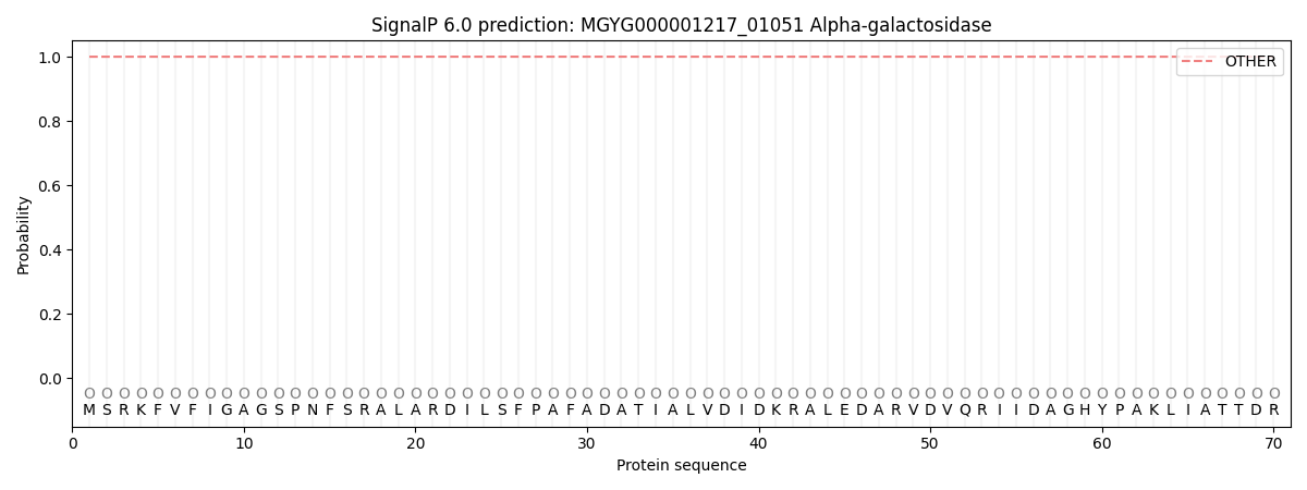
This protein is predicted as OTHER

| Other | SP_Sec_SPI | LIPO_Sec_SPII | TAT_Tat_SPI | TATLIP_Sec_SPII | PILIN_Sec_SPIII |
|---|---|---|---|---|---|
| 1.000049 | 0.000000 | 0.000000 | 0.000000 | 0.000000 | 0.000000 |
