You are browsing environment: HUMAN GUT
CAZyme Information: MGYG000001262_02296
You are here: Home > Sequence: MGYG000001262_02296
Basic Information |
Genomic context |
Full Sequence |
Enzyme annotations |
CAZy signature domains |
CDD domains |
CAZyme hits |
PDB hits |
Swiss-Prot hits |
SignalP and Lipop annotations |
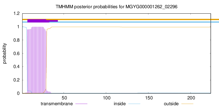
TMHMM annotations
Basic Information help
| Species | Sphingomonas ginsenosidimutans | |||||||||||
|---|---|---|---|---|---|---|---|---|---|---|---|---|
| Lineage | Bacteria; Proteobacteria; Alphaproteobacteria; Sphingomonadales; Sphingomonadaceae; Sphingomonas; Sphingomonas ginsenosidimutans | |||||||||||
| CAZyme ID | MGYG000001262_02296 | |||||||||||
| CAZy Family | GT51 | |||||||||||
| CAZyme Description | Biosynthetic peptidoglycan transglycosylase | |||||||||||
| CAZyme Property |
|
|||||||||||
| Genome Property |
|
|||||||||||
| Gene Location | Start: 12476; End: 13150 Strand: + | |||||||||||
CAZyme Signature Domains help
| Family | Start | End | Evalue | family coverage |
|---|---|---|---|---|
| GT51 | 48 | 196 | 9.8e-48 | 0.8418079096045198 |
CDD Domains download full data without filtering help
| Cdd ID | Domain | E-Value | qStart | qEnd | sStart | sEnd | Domain Description |
|---|---|---|---|---|---|---|---|
| PRK00056 | mtgA | 6.49e-91 | 3 | 202 | 7 | 214 | monofunctional biosynthetic peptidoglycan transglycosylase; Provisional |
| TIGR02070 | mono_pep_trsgly | 2.10e-73 | 4 | 216 | 3 | 223 | monofunctional biosynthetic peptidoglycan transglycosylase. This family is one of the transglycosylases involved in the late stages of peptidoglycan biosynthesis. Members tend to be small, about 240 amino acids in length, and consist almost entirely of a domain described by pfam00912 for transglycosylases. Species with this protein will have several other transglycosylases as well. All species with this protein are Proteobacteria that produce murein (peptidoglycan). [Cell envelope, Biosynthesis and degradation of murein sacculus and peptidoglycan] |
| pfam00912 | Transgly | 3.53e-57 | 50 | 216 | 14 | 175 | Transglycosylase. The penicillin-binding proteins are bifunctional proteins consisting of transglycosylase and transpeptidase in the N- and C-terminus respectively. The transglycosylase domain catalyzes the polymerization of murein glycan chains. |
| COG0744 | MrcB | 1.47e-52 | 1 | 202 | 17 | 231 | Membrane carboxypeptidase (penicillin-binding protein) [Cell wall/membrane/envelope biogenesis]. |
| TIGR02074 | PBP_1a_fam | 4.05e-39 | 51 | 190 | 4 | 144 | penicillin-binding protein, 1A family. Bacterial that synthesize a cell wall of peptidoglycan (murein) generally have several transglycosylases and transpeptidases for the task. This family consists of bifunctional transglycosylase/transpeptidase penicillin-binding proteins (PBP). In the Proteobacteria, this family includes PBP 1A but not the paralogous PBP 1B (TIGR02071). This family also includes related proteins, often designated PBP 1A, from other bacterial lineages. [Cell envelope, Biosynthesis and degradation of murein sacculus and peptidoglycan] |
CAZyme Hits help
| Hit ID | E-Value | Query Start | Query End | Hit Start | Hit End |
|---|---|---|---|---|---|
| QKS00874.1 | 1.01e-121 | 10 | 224 | 11 | 225 |
| QBE91723.1 | 8.02e-121 | 9 | 224 | 9 | 224 |
| QRY96267.1 | 8.02e-121 | 9 | 224 | 9 | 224 |
| BCI70380.1 | 8.02e-121 | 9 | 224 | 9 | 224 |
| AOW25090.1 | 1.26e-120 | 5 | 223 | 5 | 223 |
PDB Hits download full data without filtering help
| Hit ID | E-Value | Query Start | Query End | Hit Start | Hit End | Description |
|---|---|---|---|---|---|---|
| 3NB6_A | 3.72e-23 | 51 | 190 | 22 | 162 | Crystalstructure of Aquifex aeolicus peptidoglycan glycosyltransferase in complex with Methylphosphoryl Neryl Moenomycin [Aquifex aeolicus] |
| 2OQO_A | 3.72e-23 | 51 | 190 | 22 | 162 | Crystalstructure of a peptidoglycan glycosyltransferase from a class A PBP: insight into bacterial cell wall synthesis [Aquifex aeolicus VF5],3D3H_A Crystal structure of a complex of the peptidoglycan glycosyltransferase domain from Aquifex aeolicus and neryl moenomycin A [Aquifex aeolicus],3NB7_A Crystal structure of Aquifex Aeolicus Peptidoglycan Glycosyltransferase in complex with Decarboxylated Neryl Moenomycin [Aquifex aeolicus] |
| 5U2G_A | 1.43e-20 | 64 | 202 | 55 | 191 | 2.6Angstrom Resolution Crystal Structure of Penicillin-Binding Protein 1A from Haemophilus influenzae [Haemophilus influenzae Rd KW20],5U2G_B 2.6 Angstrom Resolution Crystal Structure of Penicillin-Binding Protein 1A from Haemophilus influenzae [Haemophilus influenzae Rd KW20] |
| 7U4H_A | 5.47e-17 | 54 | 188 | 45 | 178 | ChainA, Penicillin-binding protein 1A (Pbp1a) [Chlamydia trachomatis D/UW-3/CX],7U4H_B Chain B, Penicillin-binding protein 1A (Pbp1a) [Chlamydia trachomatis D/UW-3/CX] |
| 4OON_A | 1.79e-16 | 57 | 209 | 47 | 201 | Crystalstructure of PBP1a in complex with compound 17 ((4Z,8S,11E,14S)-5-(2-amino-1,3-thiazol-4-yl)-14-(5,6-dihydroxy-1,3-dioxo-1,3-dihydro-2H-isoindol-2-yl)-8-formyl-2-methyl-6-oxo-3,10-dioxa-4,7,11-triazatetradeca-4,11-diene-2,12,14-tricarboxylic acid) [Pseudomonas aeruginosa PAO1] |
Swiss-Prot Hits download full data without filtering help
| Hit ID | E-Value | Query Start | Query End | Hit Start | Hit End | Description |
|---|---|---|---|---|---|---|
| B8GYX9 | 8.40e-68 | 23 | 223 | 28 | 228 | Biosynthetic peptidoglycan transglycosylase OS=Caulobacter vibrioides (strain NA1000 / CB15N) OX=565050 GN=mtgA PE=3 SV=1 |
| Q9ABA6 | 8.40e-68 | 23 | 223 | 28 | 228 | Biosynthetic peptidoglycan transglycosylase OS=Caulobacter vibrioides (strain ATCC 19089 / CB15) OX=190650 GN=mtgA PE=3 SV=1 |
| B6IWJ6 | 3.45e-57 | 26 | 223 | 29 | 227 | Biosynthetic peptidoglycan transglycosylase OS=Rhodospirillum centenum (strain ATCC 51521 / SW) OX=414684 GN=mtgA PE=3 SV=1 |
| B0RQA5 | 6.99e-55 | 19 | 202 | 35 | 225 | Biosynthetic peptidoglycan transglycosylase OS=Xanthomonas campestris pv. campestris (strain B100) OX=509169 GN=mtgA PE=3 SV=1 |
| Q8P6V1 | 6.99e-55 | 19 | 202 | 35 | 225 | Biosynthetic peptidoglycan transglycosylase OS=Xanthomonas campestris pv. campestris (strain ATCC 33913 / DSM 3586 / NCPPB 528 / LMG 568 / P 25) OX=190485 GN=mtgA PE=3 SV=1 |
SignalP and Lipop Annotations help
This protein is predicted as OTHER
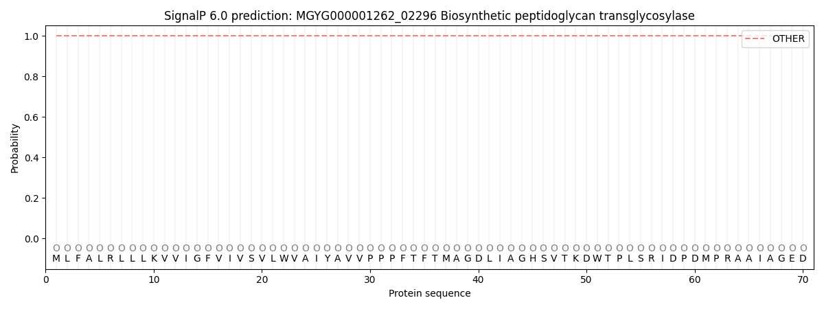
| Other | SP_Sec_SPI | LIPO_Sec_SPII | TAT_Tat_SPI | TATLIP_Sec_SPII | PILIN_Sec_SPIII |
|---|---|---|---|---|---|
| 1.000026 | 0.000002 | 0.000000 | 0.000000 | 0.000000 | 0.000000 |