You are browsing environment: HUMAN GUT
CAZyme Information: MGYG000001279_02660
You are here: Home > Sequence: MGYG000001279_02660
Basic Information |
Genomic context |
Full Sequence |
Enzyme annotations |
CAZy signature domains |
CDD domains |
CAZyme hits |
PDB hits |
Swiss-Prot hits |
SignalP and Lipop annotations |
TMHMM annotations
Basic Information help
| Species | Pseudomonas_E fulva | |||||||||||
|---|---|---|---|---|---|---|---|---|---|---|---|---|
| Lineage | Bacteria; Proteobacteria; Gammaproteobacteria; Pseudomonadales; Pseudomonadaceae; Pseudomonas_E; Pseudomonas_E fulva | |||||||||||
| CAZyme ID | MGYG000001279_02660 | |||||||||||
| CAZy Family | CBM48 | |||||||||||
| CAZyme Description | 1,4-alpha-glucan branching enzyme GlgB | |||||||||||
| CAZyme Property |
|
|||||||||||
| Genome Property |
|
|||||||||||
| Gene Location | Start: 2022; End: 4232 Strand: + | |||||||||||
CAZyme Signature Domains help
| Family | Start | End | Evalue | family coverage |
|---|---|---|---|---|
| GH13 | 286 | 586 | 8.9e-164 | 0.9966777408637874 |
| CBM48 | 128 | 214 | 5.6e-20 | 0.9078947368421053 |
CDD Domains download full data without filtering help
| Cdd ID | Domain | E-Value | qStart | qEnd | sStart | sEnd | Domain Description |
|---|---|---|---|---|---|---|---|
| PRK14706 | PRK14706 | 0.0 | 129 | 736 | 27 | 622 | glycogen branching enzyme; Provisional |
| PRK12313 | PRK12313 | 0.0 | 106 | 736 | 4 | 628 | 1,4-alpha-glucan branching protein GlgB. |
| TIGR01515 | branching_enzym | 0.0 | 113 | 734 | 1 | 618 | alpha-1,4-glucan:alpha-1,4-glucan 6-glycosyltransferase. This model describes the glycogen branching enzymes which are responsible for the transfer of chains of approx. 7 alpha(1--4)-linked glucosyl residues to other similar chains (in new alpha(1--6) linkages) in the biosynthesis of glycogen. This enzyme is a member of the broader amylase family of starch hydrolases which fold as (beta/alpha)8 barrels, the so-called TIM-barrel structure. All of the sequences comprising the seed of this model have been experimentally characterized. This model encompasses both bacterial and eukaryotic species. No archaea have this enzyme, although Aquifex aolicus does. Two species, Bacillus thuringiensis and Clostridium perfringens have two sequences each which are annotated as amylases. These annotations are aparrently in error. GP|18143720 from C. perfringens, for instance, contains the note "674 aa, similar to gp:A14658_1 amylase (1,4-alpha-glucan branching enzyme (EC 2.4.1.18) ) from Bacillus thuringiensis (648 aa); 51.1% identity in 632 aa overlap." A branching enzyme from Porphyromonas gingivales, OMNI|PG1793, appears to be more closely related to the eukaryotic species (across a deep phylogenetic split) and may represent an instance of lateral transfer from this species' host. A sequence from Arabidopsis thaliana, GP|9294564, scores just above trusted, but appears either to contain corrupt sequence or, more likely, to be a pseudogene as some of the conserved catalytic residues common to the alpha amylase family are not conserved here. [Energy metabolism, Biosynthesis and degradation of polysaccharides] |
| PRK05402 | PRK05402 | 0.0 | 13 | 736 | 3 | 724 | 1,4-alpha-glucan branching protein GlgB. |
| PRK14705 | PRK14705 | 0.0 | 16 | 735 | 508 | 1222 | glycogen branching enzyme; Provisional |
CAZyme Hits help
| Hit ID | E-Value | Query Start | Query End | Hit Start | Hit End |
|---|---|---|---|---|---|
| QPH47519.1 | 0.0 | 1 | 736 | 1 | 736 |
| QPH42456.1 | 0.0 | 1 | 736 | 1 | 736 |
| AVF56024.1 | 0.0 | 1 | 736 | 1 | 736 |
| QDC04051.1 | 0.0 | 1 | 736 | 1 | 736 |
| CRN07791.1 | 0.0 | 1 | 736 | 1 | 736 |
PDB Hits download full data without filtering help
| Hit ID | E-Value | Query Start | Query End | Hit Start | Hit End | Description |
|---|---|---|---|---|---|---|
| 1M7X_A | 1.50e-263 | 116 | 729 | 1 | 608 | TheX-ray Crystallographic Structure of Branching Enzyme [Escherichia coli],1M7X_B The X-ray Crystallographic Structure of Branching Enzyme [Escherichia coli],1M7X_C The X-ray Crystallographic Structure of Branching Enzyme [Escherichia coli],1M7X_D The X-ray Crystallographic Structure of Branching Enzyme [Escherichia coli] |
| 4LPC_A | 1.17e-261 | 130 | 729 | 10 | 603 | CrystalStructure of E.Coli Branching Enzyme in complex with maltoheptaose [Escherichia coli],4LPC_B Crystal Structure of E.Coli Branching Enzyme in complex with maltoheptaose [Escherichia coli],4LPC_C Crystal Structure of E.Coli Branching Enzyme in complex with maltoheptaose [Escherichia coli],4LPC_D Crystal Structure of E.Coli Branching Enzyme in complex with maltoheptaose [Escherichia coli],4LQ1_A Crystal Structure of E.Coli Branching Enzyme in complex with maltohexaose [Escherichia coli],4LQ1_B Crystal Structure of E.Coli Branching Enzyme in complex with maltohexaose [Escherichia coli],4LQ1_C Crystal Structure of E.Coli Branching Enzyme in complex with maltohexaose [Escherichia coli],4LQ1_D Crystal Structure of E.Coli Branching Enzyme in complex with maltohexaose [Escherichia coli],5E6Y_A Crystal structure of E.Coli branching enzyme in complex with alpha cyclodextrin [Escherichia coli O139:H28 str. E24377A],5E6Y_B Crystal structure of E.Coli branching enzyme in complex with alpha cyclodextrin [Escherichia coli O139:H28 str. E24377A],5E6Y_C Crystal structure of E.Coli branching enzyme in complex with alpha cyclodextrin [Escherichia coli O139:H28 str. E24377A],5E6Y_D Crystal structure of E.Coli branching enzyme in complex with alpha cyclodextrin [Escherichia coli O139:H28 str. E24377A],5E6Z_A Crystal structure of Ecoli Branching Enzyme with beta cyclodextrin [Escherichia coli O139:H28 str. E24377A],5E6Z_B Crystal structure of Ecoli Branching Enzyme with beta cyclodextrin [Escherichia coli O139:H28 str. E24377A],5E6Z_C Crystal structure of Ecoli Branching Enzyme with beta cyclodextrin [Escherichia coli O139:H28 str. E24377A],5E6Z_D Crystal structure of Ecoli Branching Enzyme with beta cyclodextrin [Escherichia coli O139:H28 str. E24377A],5E70_A Crystal structure of Ecoli Branching Enzyme with gamma cyclodextrin [Escherichia coli O139:H28 str. E24377A],5E70_B Crystal structure of Ecoli Branching Enzyme with gamma cyclodextrin [Escherichia coli O139:H28 str. E24377A],5E70_C Crystal structure of Ecoli Branching Enzyme with gamma cyclodextrin [Escherichia coli O139:H28 str. E24377A],5E70_D Crystal structure of Ecoli Branching Enzyme with gamma cyclodextrin [Escherichia coli O139:H28 str. E24377A] |
| 5GQW_A | 1.05e-257 | 24 | 734 | 38 | 772 | Crystalstructure of branching enzyme W610N mutant from Cyanothece sp. ATCC 51142 [Crocosphaera subtropica ATCC 51142],5GQX_A Crystal structure of branching enzyme W610N mutant from Cyanothece sp. ATCC 51142 in complex with maltoheptaose [Crocosphaera subtropica ATCC 51142] |
| 5GR5_A | 3.00e-257 | 24 | 734 | 38 | 772 | Crystalstructure of branching enzyme W610A mutant from Cyanothece sp. ATCC 51142 [Crocosphaera subtropica ATCC 51142] |
| 5GQZ_A | 4.25e-257 | 24 | 734 | 38 | 772 | Crystalstructure of branching enzyme Y500A mutant from Cyanothece sp. ATCC 51142 [Crocosphaera subtropica ATCC 51142] |
Swiss-Prot Hits download full data without filtering help
| Hit ID | E-Value | Query Start | Query End | Hit Start | Hit End | Description |
|---|---|---|---|---|---|---|
| Q4KCQ3 | 0.0 | 1 | 735 | 1 | 741 | 1,4-alpha-glucan branching enzyme GlgB OS=Pseudomonas fluorescens (strain ATCC BAA-477 / NRRL B-23932 / Pf-5) OX=220664 GN=glgB PE=3 SV=1 |
| Q5H6H2 | 0.0 | 3 | 735 | 9 | 730 | 1,4-alpha-glucan branching enzyme GlgB 1 OS=Xanthomonas oryzae pv. oryzae (strain KACC10331 / KXO85) OX=291331 GN=glgB1 PE=3 SV=1 |
| Q4ZTJ2 | 0.0 | 15 | 735 | 19 | 738 | 1,4-alpha-glucan branching enzyme GlgB OS=Pseudomonas syringae pv. syringae (strain B728a) OX=205918 GN=glgB PE=3 SV=1 |
| Q4V0E2 | 0.0 | 18 | 735 | 12 | 722 | 1,4-alpha-glucan branching enzyme GlgB 1 OS=Xanthomonas campestris pv. campestris (strain 8004) OX=314565 GN=glgB1 PE=3 SV=1 |
| Q881X0 | 0.0 | 15 | 735 | 19 | 738 | 1,4-alpha-glucan branching enzyme GlgB OS=Pseudomonas syringae pv. tomato (strain ATCC BAA-871 / DC3000) OX=223283 GN=glgB PE=3 SV=1 |
SignalP and Lipop Annotations help
This protein is predicted as OTHER
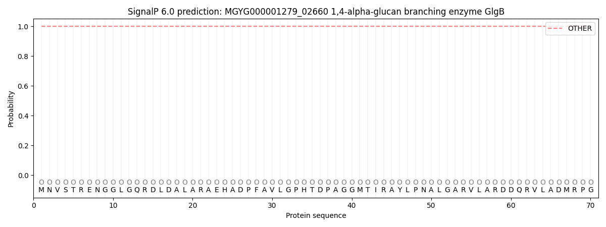
| Other | SP_Sec_SPI | LIPO_Sec_SPII | TAT_Tat_SPI | TATLIP_Sec_SPII | PILIN_Sec_SPIII |
|---|---|---|---|---|---|
| 1.000042 | 0.000002 | 0.000000 | 0.000000 | 0.000000 | 0.000000 |