You are browsing environment: HUMAN GUT
CAZyme Information: MGYG000001295_01383
You are here: Home > Sequence: MGYG000001295_01383
Basic Information |
Genomic context |
Full Sequence |
Enzyme annotations |
CAZy signature domains |
CDD domains |
CAZyme hits |
PDB hits |
Swiss-Prot hits |
SignalP and Lipop annotations |
TMHMM annotations
Basic Information help
| Species | Desulfovibrio fairfieldensis | |||||||||||
|---|---|---|---|---|---|---|---|---|---|---|---|---|
| Lineage | Bacteria; Desulfobacterota; Desulfovibrionia; Desulfovibrionales; Desulfovibrionaceae; Desulfovibrio; Desulfovibrio fairfieldensis | |||||||||||
| CAZyme ID | MGYG000001295_01383 | |||||||||||
| CAZy Family | GT4 | |||||||||||
| CAZyme Description | D-inositol-3-phosphate glycosyltransferase | |||||||||||
| CAZyme Property |
|
|||||||||||
| Genome Property |
|
|||||||||||
| Gene Location | Start: 636071; End: 637741 Strand: - | |||||||||||
CDD Domains download full data without filtering help
| Cdd ID | Domain | E-Value | qStart | qEnd | sStart | sEnd | Domain Description |
|---|---|---|---|---|---|---|---|
| cd03801 | GT4_PimA-like | 6.51e-33 | 40 | 423 | 33 | 365 | phosphatidyl-myo-inositol mannosyltransferase. This family is most closely related to the GT4 family of glycosyltransferases and named after PimA in Propionibacterium freudenreichii, which is involved in the biosynthesis of phosphatidyl-myo-inositol mannosides (PIM) which are early precursors in the biosynthesis of lipomannans (LM) and lipoarabinomannans (LAM), and catalyzes the addition of a mannosyl residue from GDP-D-mannose (GDP-Man) to the position 2 of the carrier lipid phosphatidyl-myo-inositol (PI) to generate a phosphatidyl-myo-inositol bearing an alpha-1,2-linked mannose residue (PIM1). Glycosyltransferases catalyze the transfer of sugar moieties from activated donor molecules to specific acceptor molecules, forming glycosidic bonds. The acceptor molecule can be a lipid, a protein, a heterocyclic compound, or another carbohydrate residue. This group of glycosyltransferases is most closely related to the previously defined glycosyltransferase family 1 (GT1). The members of this family may transfer UDP, ADP, GDP, or CMP linked sugars. The diverse enzymatic activities among members of this family reflect a wide range of biological functions. The protein structure available for this family has the GTB topology, one of the two protein topologies observed for nucleotide-sugar-dependent glycosyltransferases. GTB proteins have distinct N- and C- terminal domains each containing a typical Rossmann fold. The two domains have high structural homology despite minimal sequence homology. The large cleft that separates the two domains includes the catalytic center and permits a high degree of flexibility. The members of this family are found mainly in certain bacteria and archaea. |
| cd03808 | GT4_CapM-like | 7.06e-24 | 285 | 418 | 255 | 356 | capsular polysaccharide biosynthesis glycosyltransferase CapM and similar proteins. This family is most closely related to the GT4 family of glycosyltransferases. CapM in Staphylococcus aureus is required for the synthesis of type 1 capsular polysaccharides. |
| COG0438 | RfaB | 1.67e-23 | 89 | 426 | 87 | 377 | Glycosyltransferase involved in cell wall bisynthesis [Cell wall/membrane/envelope biogenesis]. |
| cd03820 | GT4_AmsD-like | 4.23e-20 | 282 | 421 | 244 | 351 | amylovoran biosynthesis glycosyltransferase AmsD and similar proteins. This family is most closely related to the GT4 family of glycosyltransferases. AmSD in Erwinia amylovora has been shown to be involved in the biosynthesis of amylovoran, the acidic exopolysaccharide acting as a virulence factor. This enzyme may be responsible for the formation of galactose alpha-1,6 linkages in amylovoran. |
| pfam00534 | Glycos_transf_1 | 2.04e-18 | 285 | 405 | 70 | 158 | Glycosyl transferases group 1. Mutations in this domain of PIGA lead to disease (Paroxysmal Nocturnal haemoglobinuria). Members of this family transfer activated sugars to a variety of substrates, including glycogen, Fructose-6-phosphate and lipopolysaccharides. Members of this family transfer UDP, ADP, GDP or CMP linked sugars. The eukaryotic glycogen synthases may be distant members of this family. |
CAZyme Hits help
| Hit ID | E-Value | Query Start | Query End | Hit Start | Hit End |
|---|---|---|---|---|---|
| AMD91220.1 | 0.0 | 1 | 556 | 1 | 556 |
| ABM28374.1 | 3.48e-143 | 5 | 522 | 16 | 523 |
| ADP86654.1 | 1.96e-142 | 5 | 522 | 16 | 523 |
| AAS96281.1 | 1.96e-142 | 5 | 522 | 16 | 523 |
| QLA16151.1 | 3.40e-142 | 5 | 554 | 6 | 560 |
PDB Hits download full data without filtering help
| Hit ID | E-Value | Query Start | Query End | Hit Start | Hit End | Description |
|---|---|---|---|---|---|---|
| 3C4Q_A | 2.50e-09 | 163 | 364 | 179 | 372 | Structureof the retaining glycosyltransferase MshA : The first step in mycothiol biosynthesis. Organism : Corynebacterium glutamicum- Complex with UDP [Corynebacterium glutamicum],3C4Q_B Structure of the retaining glycosyltransferase MshA : The first step in mycothiol biosynthesis. Organism : Corynebacterium glutamicum- Complex with UDP [Corynebacterium glutamicum],3C4V_A Structure of the retaining glycosyltransferase MshA:The first step in mycothiol biosynthesis. Organism: Corynebacterium glutamicum : Complex with UDP and 1L-INS-1-P. [Corynebacterium glutamicum],3C4V_B Structure of the retaining glycosyltransferase MshA:The first step in mycothiol biosynthesis. Organism: Corynebacterium glutamicum : Complex with UDP and 1L-INS-1-P. [Corynebacterium glutamicum] |
| 3C48_A | 2.58e-09 | 163 | 364 | 199 | 392 | Structureof the retaining glycosyltransferase MshA: The first step in mycothiol biosynthesis. Organism: Corynebacterium glutamicum- APO (OPEN) structure. [Corynebacterium glutamicum],3C48_B Structure of the retaining glycosyltransferase MshA: The first step in mycothiol biosynthesis. Organism: Corynebacterium glutamicum- APO (OPEN) structure. [Corynebacterium glutamicum] |
| 3OKA_A | 3.79e-09 | 176 | 413 | 163 | 366 | Crystalstructure of Corynebacterium glutamicum PimB' in complex with GDP-Man (triclinic crystal form) [Corynebacterium glutamicum],3OKA_B Crystal structure of Corynebacterium glutamicum PimB' in complex with GDP-Man (triclinic crystal form) [Corynebacterium glutamicum] |
| 3OKC_A | 3.97e-09 | 176 | 413 | 163 | 366 | Crystalstructure of Corynebacterium glutamicum PimB' bound to GDP (orthorhombic crystal form) [Corynebacterium glutamicum],3OKP_A Crystal structure of Corynebacterium glutamicum PimB' bound to GDP-Man (orthorhombic crystal form) [Corynebacterium glutamicum] |
Swiss-Prot Hits download full data without filtering help
| Hit ID | E-Value | Query Start | Query End | Hit Start | Hit End | Description |
|---|---|---|---|---|---|---|
| D2S4K7 | 4.32e-14 | 162 | 350 | 191 | 373 | D-inositol 3-phosphate glycosyltransferase OS=Geodermatophilus obscurus (strain ATCC 25078 / DSM 43160 / JCM 3152 / KCC A-0152 / KCTC 9177 / NBRC 13315 / NRRL B-3577 / G-20) OX=526225 GN=mshA PE=3 SV=1 |
| C8XA09 | 7.87e-13 | 152 | 443 | 193 | 441 | D-inositol 3-phosphate glycosyltransferase OS=Nakamurella multipartita (strain ATCC 700099 / DSM 44233 / CIP 104796 / JCM 9543 / NBRC 105858 / Y-104) OX=479431 GN=mshA PE=3 SV=1 |
| D3Q051 | 1.24e-11 | 167 | 449 | 205 | 443 | D-inositol 3-phosphate glycosyltransferase OS=Stackebrandtia nassauensis (strain DSM 44728 / CIP 108903 / NRRL B-16338 / NBRC 102104 / LLR-40K-21) OX=446470 GN=mshA PE=3 SV=1 |
| C3PK12 | 2.66e-11 | 163 | 412 | 179 | 391 | D-inositol 3-phosphate glycosyltransferase OS=Corynebacterium aurimucosum (strain ATCC 700975 / DSM 44827 / CIP 107346 / CN-1) OX=548476 GN=mshA PE=3 SV=1 |
| A6W6D9 | 8.72e-11 | 151 | 412 | 172 | 408 | D-inositol 3-phosphate glycosyltransferase OS=Kineococcus radiotolerans (strain ATCC BAA-149 / DSM 14245 / SRS30216) OX=266940 GN=mshA PE=3 SV=1 |
SignalP and Lipop Annotations help
This protein is predicted as OTHER
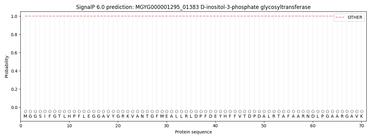
| Other | SP_Sec_SPI | LIPO_Sec_SPII | TAT_Tat_SPI | TATLIP_Sec_SPII | PILIN_Sec_SPIII |
|---|---|---|---|---|---|
| 1.000057 | 0.000000 | 0.000000 | 0.000000 | 0.000000 | 0.000000 |