You are browsing environment: HUMAN GUT
CAZyme Information: MGYG000001306_03295
You are here: Home > Sequence: MGYG000001306_03295
Basic Information |
Genomic context |
Full Sequence |
Enzyme annotations |
CAZy signature domains |
CDD domains |
CAZyme hits |
PDB hits |
Swiss-Prot hits |
SignalP and Lipop annotations |
TMHMM annotations
Basic Information help
| Species | Phocaeicola coprocola | |||||||||||
|---|---|---|---|---|---|---|---|---|---|---|---|---|
| Lineage | Bacteria; Bacteroidota; Bacteroidia; Bacteroidales; Bacteroidaceae; Phocaeicola; Phocaeicola coprocola | |||||||||||
| CAZyme ID | MGYG000001306_03295 | |||||||||||
| CAZy Family | GH20 | |||||||||||
| CAZyme Description | hypothetical protein | |||||||||||
| CAZyme Property |
|
|||||||||||
| Genome Property |
|
|||||||||||
| Gene Location | Start: 69123; End: 69980 Strand: + | |||||||||||
CAZyme Signature Domains help
| Family | Start | End | Evalue | family coverage |
|---|---|---|---|---|
| GH20 | 146 | 273 | 6.4e-38 | 0.36795252225519287 |
CDD Domains download full data without filtering help
| Cdd ID | Domain | E-Value | qStart | qEnd | sStart | sEnd | Domain Description |
|---|---|---|---|---|---|---|---|
| cd06564 | GH20_DspB_LnbB-like | 1.60e-51 | 153 | 283 | 1 | 137 | Glycosyl hydrolase family 20 (GH20) catalytic domain of dispersin B (DspB), lacto-N-biosidase (LnbB) and related proteins. Dispersin B is a soluble beta-N-acetylglucosamidase found in bacteria that hydrolyzes the beta-1,6-linkages of PGA (poly-beta-(1,6)-N-acetylglucosamine), a major component of the extracellular polysaccharide matrix. Lacto-N-biosidase hydrolyzes lacto-N-biose (LNB) type I oligosaccharides at the nonreducing terminus to produce lacto-N-biose as part of the GNB/LNB (galacto-N-biose/lacto-N-biose I) degradation pathway. The lacto-N-biosidase from Bifidobacterium bifidum has this GH20 domain, a carbohydrate binding module 32, and a bacterial immunoglobulin-like domain 2, as well as a YSIRK signal peptide and a G5 membrane anchor at the N and C termini, respectively. The GH20 hexosaminidases are thought to act via a catalytic mechanism in which the catalytic nucleophile is not provided by solvent or the enzyme, but by the substrate itself. |
| pfam02838 | Glyco_hydro_20b | 1.79e-28 | 24 | 148 | 2 | 123 | Glycosyl hydrolase family 20, domain 2. This domain has a zincin-like fold. |
| pfam00728 | Glyco_hydro_20 | 3.57e-26 | 152 | 258 | 1 | 106 | Glycosyl hydrolase family 20, catalytic domain. This domain has a TIM barrel fold. |
| cd06563 | GH20_chitobiase-like | 8.70e-24 | 152 | 258 | 1 | 114 | The chitobiase of Serratia marcescens is a beta-N-1,4-acetylhexosaminidase with a glycosyl hydrolase family 20 (GH20) domain that hydrolyzes the beta-1,4-glycosidic linkages in oligomers derived from chitin. Chitin is degraded by a two step process: i) a chitinase hydrolyzes the chitin to oligosaccharides and disaccharides such as di-N-acetyl-D-glucosamine and chitobiose, ii) chitobiase then further degrades these oligomers into monomers. This GH20 domain family includes an N-acetylglucosamidase (GlcNAcase A) from Pseudoalteromonas piscicida and an N-acetylhexosaminidase (SpHex) from Streptomyces plicatus. SpHex lacks the C-terminal PKD (polycystic kidney disease I)-like domain found in the chitobiases. The GH20 hexosaminidases are thought to act via a catalytic mechanism in which the catalytic nucleophile is not provided by solvent or the enzyme, but by the substrate itself. |
| cd06568 | GH20_SpHex_like | 2.06e-23 | 155 | 271 | 4 | 116 | A subgroup of the Glycosyl hydrolase family 20 (GH20) catalytic domain found in proteins similar to the N-acetylhexosaminidase from Streptomyces plicatus (SpHex). SpHex catalyzes the hydrolysis of N-acetyl-beta-hexosaminides. An Asp residue within the active site plays a critical role in substrate-assisted catalysis by orienting the 2-acetamido group and stabilizing the transition state. The GH20 hexosaminidases are thought to act via a catalytic mechanism in which the catalytic nucleophile is not provided by solvent or the enzyme, but by the substrate itself. Proteins belonging to this subgroup lack the C-terminal PKD (polycystic kidney disease I)-like domain found in the chitobiases. |
CAZyme Hits help
| Hit ID | E-Value | Query Start | Query End | Hit Start | Hit End |
|---|---|---|---|---|---|
| SCV07807.1 | 8.88e-141 | 2 | 283 | 3 | 284 |
| ALJ48397.1 | 8.88e-141 | 2 | 283 | 3 | 284 |
| QRQ55239.1 | 8.88e-141 | 2 | 283 | 3 | 284 |
| QGT73121.1 | 1.26e-140 | 2 | 283 | 3 | 284 |
| QNL40659.1 | 1.78e-140 | 2 | 283 | 3 | 284 |
PDB Hits download full data without filtering help
| Hit ID | E-Value | Query Start | Query End | Hit Start | Hit End | Description |
|---|---|---|---|---|---|---|
| 6JQF_A | 4.61e-43 | 24 | 266 | 89 | 333 | Crystallizationanalysis of a beta-N-acetylhexosaminidase (Am2136) from Akkermansia muciniphila [Akkermansia muciniphila ATCC BAA-835] |
| 3GH4_A | 1.63e-25 | 24 | 275 | 39 | 301 | Crystalstructure of beta-hexosaminidase from Paenibacillus sp. TS12 [Paenibacillus sp.],3GH5_A Crystal structure of beta-hexosaminidase from Paenibacillus sp. TS12 in complex with GlcNAc [Paenibacillus sp.],3GH7_A Crystal structure of beta-hexosaminidase from Paenibacillus sp. TS12 in complex with GalNAc [Paenibacillus sp.],3SUR_A Crystal structure of beta-hexosaminidase from Paenibacillus sp. TS12 in complex with NAG-thiazoline. [Paenibacillus sp. TS12],3SUS_A Crystal structure of beta-hexosaminidase from Paenibacillus sp. TS12 in complex with Gal-NAG-thiazoline [Paenibacillus sp. TS12],3SUT_A Crystal structure of beta-hexosaminidase from Paenibacillus sp. TS12 in complex with PUGNAc [Paenibacillus sp. TS12],3SUU_A Crystal structure of beta-hexosaminidase from Paenibacillus sp. TS12 in complex with Gal-PUGNAc [Paenibacillus sp. TS12],3SUV_A Crystal structure of beta-hexosaminidase from Paenibacillus sp. TS12 in complex with NHAc-DNJ [Paenibacillus sp. TS12],3SUW_A Crystal structure of beta-hexosaminidase from Paenibacillus sp. TS12 in complex with NHAc-CAS [Paenibacillus sp. TS12] |
| 6Q63_A | 5.12e-24 | 6 | 273 | 13 | 287 | BT0459[Bacteroides thetaiotaomicron],6Q63_B BT0459 [Bacteroides thetaiotaomicron],6Q63_C BT0459 [Bacteroides thetaiotaomicron] |
| 4C7D_A | 1.29e-22 | 25 | 258 | 8 | 241 | Structureand activity of the GH20 beta-N-acetylhexosaminidase from Streptomyces coelicolor A3(2) [Streptomyces coelicolor],4C7D_B Structure and activity of the GH20 beta-N-acetylhexosaminidase from Streptomyces coelicolor A3(2) [Streptomyces coelicolor],4C7F_A Structure and activity of the GH20 beta-N-acetylhexosaminidase from Streptomyces coelicolor A3(2) [Streptomyces coelicolor],4C7F_B Structure and activity of the GH20 beta-N-acetylhexosaminidase from Streptomyces coelicolor A3(2) [Streptomyces coelicolor] |
| 4C7G_A | 1.29e-22 | 25 | 258 | 8 | 241 | Structureand activity of the GH20 beta-N-acetylhexosaminidase from Streptomyces coelicolor A3(2) [Streptomyces coelicolor] |
Swiss-Prot Hits download full data without filtering help
| Hit ID | E-Value | Query Start | Query End | Hit Start | Hit End | Description |
|---|---|---|---|---|---|---|
| B2UPR7 | 3.68e-45 | 24 | 266 | 111 | 355 | Beta-hexosaminidase Amuc_2136 OS=Akkermansia muciniphila (strain ATCC BAA-835 / DSM 22959 / JCM 33894 / BCRC 81048 / CCUG 64013 / CIP 107961 / Muc) OX=349741 GN=Amuc_2136 PE=1 SV=1 |
| P49008 | 6.48e-21 | 26 | 273 | 37 | 289 | Beta-hexosaminidase OS=Porphyromonas gingivalis (strain ATCC BAA-308 / W83) OX=242619 GN=nahA PE=3 SV=2 |
| B2UP57 | 7.05e-20 | 83 | 258 | 37 | 217 | Beta-hexosaminidase Amuc_2018 OS=Akkermansia muciniphila (strain ATCC BAA-835 / DSM 22959 / JCM 33894 / BCRC 81048 / CCUG 64013 / CIP 107961 / Muc) OX=349741 GN=Amuc_2018 PE=1 SV=1 |
| Q7WUL4 | 9.24e-18 | 88 | 261 | 68 | 240 | Beta-N-acetylhexosaminidase OS=Cellulomonas fimi OX=1708 GN=hex20 PE=1 SV=1 |
| P13723 | 1.81e-17 | 18 | 273 | 16 | 266 | Beta-hexosaminidase subunit A1 OS=Dictyostelium discoideum OX=44689 GN=hexa1 PE=1 SV=1 |
SignalP and Lipop Annotations help
This protein is predicted as SP
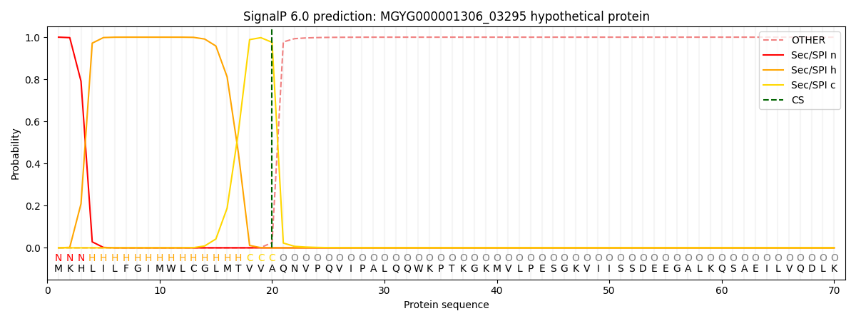
| Other | SP_Sec_SPI | LIPO_Sec_SPII | TAT_Tat_SPI | TATLIP_Sec_SPII | PILIN_Sec_SPIII |
|---|---|---|---|---|---|
| 0.000331 | 0.998944 | 0.000220 | 0.000164 | 0.000158 | 0.000153 |